|Articles|February 2, 2018
- BioPharm International-02-01-2018
- Volume 31
- Issue 2
Impurity Testing of Biologic Drug Products
Author(s)Adeline Siew, PhD
Experts share insights on the various methods used for purity and impurity analysis of therapeutic proteins.
Advertisement
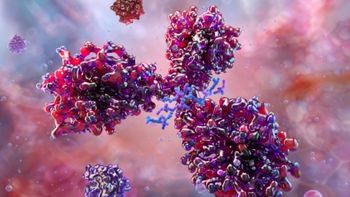
Impurities can have a negative impact on the stability, safety, and efficacy of protein therapeutics. “Aggregates are of particular concern, either in soluble dimer/oligomer form or subvisible particle form,” notes Jay Kang, director of analytical and formulation development at Patheon, part of Thermo Fisher Scientific. “It is well documented that even a small amount of aggregates can cause a significant and sometimes life-threatening immunogenic reaction.”
“Impurities may interact with the therapeutic protein in a way that blocks and/or compromises the activity and potency of the therapeutic protein in vivo, hence, reducing its efficacy,” explain Michael Sadick, principal scientist, Catalent Biologics Analytical Services, and Michael Merges director of strategic growth, Catalent Biologics. “On the other hand, the impurity may exaggerate or enhance the therapeutic protein’s bioactivity in an uncontrolled way, leading to adverse events. Some impurities (especially host cell proteins) may add an immune-stimulating or adjuvant behavior to the therapeutic, causing the patient to generate antibodies or cell-mediated immunity against the protein.”
“Host cell proteins co-extracted with the therapeutic protein can contain enzymes such as oxidases and lipases that will break down the protein over time, affecting the stability of the product,” adds Niall Dinwoodie, Edinburgh biologics site lead at Charles River Labs (CRL). “Other host cell proteins and binding agents carried over from purification columns may mimic the action of the therapeutic protein in assays, leading to mis-formulation of the product outside the therapeutic window.”
According to Dinwoodie, some impurities have less insidious effects, but can still render the therapeutic unacceptable. “For example, trace levels of rapidly oxidized materials cause significant color change,” he says. “Historically, contamination with trace amounts of metals was a problem leading to aggregation of therapeutic proteins, which can cause significant changes in efficacy, but understanding of the phenomena has led to improved, metal-free production processes.”
In general, impurities come from two major sources, observes Bérangère Tissot, general manager, SGS Life Sciences, West Chester, Pennsylvania: product-related impurities and process-related impurities. “Product-related impurities can be categorized as product variants, and basically correspond to any undesired modification of the protein amino-acid sequence or post-translational modifications,” she highlights. “Variants also include forms of the therapeutic proteins in solution that are different from the intended drug product (i.e., different conformation or aggregation state). They can also be identified as a sub-form of the therapeutic protein, possessing a biological activity either higher or lower than the one of the drug product.”
The second type of impurities are mostly related to the production processes, says Tissot. Dinwoodie adds that the materials used in the production process to support cell growth, extract, and purify the therapeutic protein must be removed from the final dosage form. “Residual amounts of these materials can be carried through the production process to become impurities in the final form,” he points out. “Examples include growth selection agents, surfactants, purification column binding agents, and viral inactivation agents.”
“The use of cells and growth media in the production process also presents risks of adventitious agents, such as viruses, entering the production system,” says Dinwoodie. “Whilst removal or inactivation of these agents is considered under the production process validation as a safety issue rather than tested in the final product as a quality concern, these agents can also be considered impurities in the product.”
Process- and product-related impurities should be carefully monitored and controlled in the production of therapeutic proteins. In this roundtable discussion, industry experts share insights on the various methods used for purity and impurity analysis of therapeutic proteins.
Method development and validation
BioPharm: What is the right approach to method development and validation for therapeutic proteins?
Kang (Patheon): Two concepts are key to approaching method development and validation for therapeutic proteins: ‘fit-for-purpose’ and ‘phase appropriate.’ A ‘fit-for-purpose’ strategy means a method should be suitable for its intended use and phase of development. The requirement to establish an analytical method depends on whether it is for an identity test, content test, or purity/impurity test; whether it is for release, in-process testing, or characterization. For example, the only requirement for an identity test is specificity, while specificity, linearity, range, precision, robustness, and sensitivity are mandatory for the purity test. Determining whether the method is for early-phase development or for biological license application (BLA) filing is also crucial because it will dictate the size and thoroughness of the validation data package.
Sadick and Merges (Catalent): The underpinning to this response is the knowledge that each protein therapeutic is quite different from any other protein therapeutic, whether in terms of its final tertiary or quaternary folding, biological activity, or purity profile. This is true even when considering different monoclonal antibody therapeutics. Consequently, while similar strategies may be used for different protein therapeutics, true ‘toolbox’ approaches/platforms may not be completely successful. In the strategies for assay or method development and optimization, we consider a combination of ‘one factor at a time’ (OFAT) to define individual factors, at least initially, and ‘design of experimentation’ (DOE) to look at multiple and interacting factors. The use of fractional factorial DOE as soon as is practical allows for a more rapid and robust method development.
Validation would be accomplished in a phase-appropriate manner. The guideline for all phase appropriate levels would be the International Council for Harmonization (ICH) Q2 (R1) (1), although different technical platforms (e.g., enzyme-linked immunosorbent assay [ELISA] or bioassay potency tests) may have specific levels of adherence to the ICH guidelines, in addition to other guidelines, for example United States Pharmacopeia General Chapters <1033> and <1034> (2, 3). Validation for an investigational new drug or at Phase I level would have the more basic requirements, with fewer tests to be executed, a smaller number of repetitions, and wider acceptance criteria. Late-phase (Phase III/BLA-enabling) validation will include all appropriate test categories, as well as robustness, with an increased number of sample repetitions along with more stringent acceptance criteria. The establishment of appropriate validation acceptance criteria should be based upon data-driven decision. Those data are best generated via a prevalidation exercise conducted prior to the drafting of each phase-appropriate validation protocol.
Tissot (SGS): This is not straightforward, as validation approaches will depend on the nature of the method, its intended use, the development stage of the product, and the type of therapeutic proteins. In all cases, the method should be evaluated, prior to its validation, through a risk management process that will dictate which parameters to validate, which acceptance criteria to aim at, and all other necessary components of a validation study. These considerations include nature and number of replicates for each of the parameters, robustness conditions, and intermediate precision details among others.
Dinwoodie (CRL): The extent of development needed for a new analytical method will depend on the purpose of the method and the body of knowledge available on the product to which it will be applied. Physicochemical methods can be largely based on compendial procedures and require little development, and parameters for platform techniques such as size-exclusion high-performance liquid chromatography (SE–HPLC) can be established from an understanding of the protein’s molecular weight.
Binding or potency assays, however, require the selection of suitable antibodies or modification of detector cell lines. Non-therapeutic host cell proteins can also present a considerable challenge to method development in ensuring that the polyclonal sera used provides full coverage of the range of proteins that may be extracted from the production cell line.
Method development also must consider the robustness of the approach and ensure that reagents and consumable items, such as columns, are readily available and consistent in the results they generate. Validation of the method will then serve to confirm the robustness of these elements and assess the variability introduced by different analysts and equipment.
Both the number of replicates run for the determination of repeatability and intermediate precision, and the number of batches of the product tested in each run are affected by the product’s stage of development. They also must be defendable in covering all possible options for how the method will be used in the future. Other aspects of method validation are more easily derived from the guidance given in ICH Q2 (R1).
Analytical methods
BioPharm: What are the commonly used analytical methods for characterizing therapeutic proteins?
Tissot (SGS): The main document used by anyone characterizing a therapeutic protein remains the ICH Q6B guidelines (4). Now these guidelines are a little outdated, mainly with regards to biophysical methods but they still remain a good basis for the design of a characterization method panel. There are many ways to address some of the key elements that need to be evaluated during a characterization study, but some of the most commonly used are listed in the following:
Physicochemical characterization:
- Liquid chromatography–tandem mass spectrometry (LC–MS/MS) following digestion for primary amino-acid sequencing, which could be completed by N-terminal sequencing using Edman degradation. The same type of methodology can be applied to the evaluation of the most common post-translational modifications
- Liquid chromatography–mass spectrometry (LC–MS) or electrospray ionization–mass spectrometry (ESI–MS) for intact molecular weight when the therapeutic protein does not present any major challenge for ionization (such as heavily glycosylated proteins)
- Amino-acid analysis and extinction coefficient estimation
- A combination of matrix-assisted laser desorption ionization–time of flight mass spectrometry (MALDI–TOF MS), LC–MS, and other liquid chromatography with ultraviolet detection (LC–UV) or high-pressure anion exchange chromatography coupled to pulsed amperometric detection (HPAEC-PAD) methods for the quantitative and qualitative analyses of N- and O-glycosylation
- Liquid chromatography or electrophoresis methodologies to evaluate product heterogeneity (charge variants, size variants, hydrophobicity variants etc.)
- Circular dichroism (CD), Fourier transform infrared spectroscopy (FTIR), intrinsic/extrinsic fluorescence for the analyses of secondary and tertiary structures
- Sedimentation velocity analytical ultracentrifugation (SV–AUC), size exclusion chromatography coupled to multi angle light scattering (SEC–MALS) or dynamic light scattering (DLS) for the analysis of quaternary structures.
Activity characterization:
- ELISA-based bioassays
- Cell-based bioassays
- Surface plasmon resonance (SPR) or bilayer interferometry (BLI) for binding activity.
Then we have to consider what are now called the emerging techniques, at least for their application to complex biologics in an industry context, which in a couple of years will become as common as the techniques previously listed. These techniques include:
- Hydrogen-deuterium exchange–mass spectrometry (HDX–MS), ion mobility-mass spectrometry, and nuclear magnetic resonance (NMR)
- Native mass spectrometry.
Sadick and Merges (Catalent): As protein therapeutics commonly have complex structures and are generally produced and/or modified by the host cell in several functional variations, analyses of these molecules require an orthogonal approach with multiple analytic modalities.
Process-related variants can be identified, quantified, and differentiated from process-related impurities of cellular origin via techniques such as SEC–HPLC, hydrophobic interaction chromatography (HIC), ion-exchange HPLC, and isoelectric focusing (IEF) capillary electrophoresis. Process-related impurities and residuals such as Protein A can be detected and quantified with ELISA assays, whereas host cell residual DNA can be quantified via quantitative polymerase chain reaction (qPCR) assays. Functional activity of the protein therapeutic can be assessed and quantified with cell-based bioassays, or, in some cases, ELISA potency assays. Process-related variants and impurities may then be more fully identified and defined using mass spectrometry-dependent analyses. Host cell derived residual protein may be assessed and identified with a combination of ELISA assays (commercial assays at early phases, and then custom assays at later phase), one-dimensional and two-dimensional Western blot analyses, and more recently, MS-based analyses.
Kang (Patheon): To characterize a protein, we need to understand its content, primary and higher order structure, potency, heterogeneity, purity, and impurity. The commonly used analytical methods for characterizing these proteins include UV spectroscopy for concentration; SEC, analytical ultracentrifugation (AUC), and field flow fractionation (FFF) for aggregate measurement; capillary gel electrophoresis (CGE) for fragment measurements; capillary isoelectric focusing (cIEF) for charge heterogeneity, biochemical/cell-based assay for potency measurement; mass spectroscopy for primary structure; FTIR, CD, or HDX for higher order structure.
Testing for impurities
BioPharm: How do you ensure that the final drug product is free from impurities that affect safety and efficacy?
Dinwoodie (CRL): Designing the production process to minimize the materials introduced during manufacturing, as well as installing appropriate purification steps, are simple sounding methods for ensuring a final drug product is impurity free. In practice, additives are required for cell growth, non-target proteins will be extracted, and downstream processing will occur, so purification steps are paramount. Control of these steps must be demonstrated by validation and/or quality control checks on the bulk drug substance. Control of impurities that could arise from the fill/finish process are then assessed for the final product.
Kang (Patheon): It is very challenging to completely remove all the impurities, but the industry can make sure that the level of impurities in the final drug product are at a safe and consistent level. A key factor to ensuring this is to develop a sensitive and robust analytical method, so all the impurities can be accurately measured and the impurity-removing capability of the downstream process can be demonstrated. For example, ELISA is the gold standard and work horse for host cell protein measurement, but it only measures the total HCP and can’t give detailed information on the level of each individual host cell protein. Mass spectroscopy can fill the gap, and is, therefore, an excellent supplemental method for host cell protein analysis.
Sadick and Merges (Catalent): A ‘pure’ protein is one that is free from any quantifiable amounts of impurities, so implementing several orthogonal methods together is necessary to assure this is the case. The complex structural properties of the protein, the nature of the potential contaminants (host cells, viruses, genetic variants, purification process), and the accuracy and appropriateness of any one given method all influence the selection of the methods used to perform the purity/impurity analysis. A subset of these analyses is executed during the purification process to assure that each purification step is performing as expected/required. The full panel is performed upon both the drug substance and the final drug product. In this way, effectiveness of and purity at each stage of processing is evaluated and assured.
Tissot (SGS): Having a product entirely free of impurities is a very arduous task, if achievable at all.
For process-related impurities, control procedures to follow the clearance of some of the process-related impurities are designed during the very early stage of the finalization of the manufacturing processes, and are refined as the processes are locked down. The use of ultra-sensitive mass spectrometry has been increasing in that very particular field, offering a greater ability to monitor such small molecule impurities at a parts per million to parts per billion level. Such methods are also commonly validated as either process-validation-related methods or even product-release methods.
For product-related impurities, the pre-IND or equivalent panel of assays, at the very early stage of the product development, includes some of the methods that will be further refined to monitor these impurities. Complementary chromatographic and electrophoretic methods using UV detection have been used to monitor therapeutic protein variants for decades, but these methods are on the verge of being replaced by multi-attribute methods (MAMs) using primarily mass spectrometry as a detection tool. The ability to not only monitor but characterize several of these variants or impurities using a single LC-MS or LC-MS/MS method will not only bring to this field more discrimination power but it is also expected to decrease the level of detection for these undesired components.
BioPharm: What are the analytical methods used for purity and impurity analysis of therapeutic proteins?
Dinwoodie (CRL): The analytical methods used to determine the levels of impurities within a therapeutic protein are those that have both the discriminatory power to separate the impurities and the sensitivity to detect and quantify low levels of the analytes. For impurities that are not closely related in structure to the therapeutic agent, such as surfactants, for example, the method can use this difference to maximize the sensitivity. Analysis of these impurities will include steps to remove all proteinaceous material to maximize the signal for charged aerosol detection or alternative measures. Sequence and glycosylation variants are closely related, or even part of the therapeutic protein; therefore, they require highly discriminatory techniques for their quantification. Capillary electrophoresis and HPLC or ultra-high-performance liquid chromatography (UHPLC) are commonly applied to resolving these variants from the more common form of the protein. Aggregates are readily separated by SE–HPLC when the aggregation is robust. Less stressful techniques such as analytical ultracentrifugation may be required where the aggregation is more fragile. For non-therapeutic host cell proteins, cell-line specific ELISA are often used though mass spectrometry techniques can provide the discriminatory and quantification power required for these complex mixtures of impurities.
References
1. ICH, Q2 (R1) Validation of Analytical Procedures: Text and Methodology, Step 4 version (1996).
2. USP, Chapter <1033>, “Biological Assay Validation,” USP 35–NF30 (US Pharmacopeial Convention, Rockville, MD, 2011).
3. USP, Chapter <1034>, “Analysis of Biological Assays,” USP 35–NF30 (US Pharmacopeial Convention, Rockville, MD, 2011).
4. ICH, Q6B Specifications: Test Procedures and Acceptance Criteria for Biotechnological/Biological Products, Step 5 (1999).
Article DetailsBioPharm International
Vol. 31, No. 2
February 2018
Page: 14–19
Citation
When referring to this article, please cite it as A. Siew, “Impurity Testing,” BioPharm International 31 (2) 2018.
Articles in this issue
about 8 years ago
The Ins and Outs of LC-Based Analytical Tools and Techniquesabout 8 years ago
Tabletop Peristaltic Liquid-Filling Machineabout 8 years ago
USB Data Logger for Temperature-Sensitive Productsabout 8 years ago
Maintaining Lab Data Integrityabout 8 years ago
Navigating Data Integrity in the Modern Lababout 8 years ago
Designing a Single-Use Biopharmaceutical Processabout 8 years ago
Container Closures: Leaving Nothing to Chanceabout 8 years ago
A New Business Model for Pharma?about 8 years ago
FDA Framework Spurs Advanced Therapiesabout 8 years ago
Opportunities and Obstacles for Generic DrugsNewsletter
Stay at the forefront of biopharmaceutical innovation—subscribe to BioPharm International for expert insights on drug development, manufacturing, compliance, and more.
Advertisement
Related Content


Advertisement
Advertisement
Trending on BioPharm International
1
How CDMO Alliances Can Provide End-to-End Service that Reduces Drug Development Time and Costs
2
FDA Clears PharmaResearch IND for Nano-Based Cancer Drug PRD-101
3
First-in-Human Study Validates Safety of Next-Generation mRNA–LNP Platform
4
High-Dose Nusinersen Slows Neurodegeneration in SMA Patients, Study Shows
5