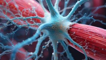
Drug-Delivering Capsules Support Transplanted Insulin-Producing Cells
Researchers at the University of Illinois developed a method to transplant pancreatic islet cells from pigs more easily to treat type I diabetes.
An in vitro study by engineers at the University of Illinois found that insulin-secreting cells called islets showed increased viability and function after spending 21 days inside small capsules containing even smaller capsules bearing a drug that makes the cells more resilient to oxygen deprivation.
The results,
Researchers have been exploring ways to transplant pancreatic islets to treat type I diabetes long term, eliminating the need for continual glucose monitoring and insulin injections. However, there are a number of challenges to this approach, according to university researchers in a Feb. 12, 2018 press release.
“First, you need viable islets that are also functional, so that they secrete insulin when exposed to glucose,” said university electrical and computer engineering professor Kyekyoon “Kevin” Kim, leader of the study. Islets from humans are scarce, he added, but pig tissue is in abundant supply, and pig insulin has been used to treat diabetes since the 1920s.
Once islets are isolated from tissue, the next big challenge is to keep them alive and functioning after transplantation. To keep the transplanted cells from interacting with the recipient’s immune system, they are packaged in small, semi-permeable capsules. The capsule size and porosity are important to allow oxygen and nutrients to reach the islets while keeping out immune cells.
“The first few weeks after transplant are very crucial because these islets need oxygen and nutrients, but do not have blood vessels to provide them,” said Hyungsoo Choi, study co-leader and senior research scientist in electrical and computer engineering at the university, in the press release. “Most critically, lack of oxygen is very toxic. It’s called hypoxia, and that will destroy the islets.”
Biological applications
Kim and Choi have developed methods of making such microcapsules for various engineering applications and realized they could use the same techniques to make microcapsules for biological applications, including drug delivery and cell transplants. Their method allows them to use materials of high viscosity, to precisely control the size and aspect ratio of the capsules, and to produce uniformly-sized microcapsules with high throughput.
“For a typical patient you’d need about 2 million capsules. Production with any other method we know cannot meet that demand easily. We’ve demonstrated that we can produce 2 million capsules in a matter of 20 minutes or so,” Kim said.
With such control and high production capacity, the researchers were able to make tiny microspheres that are loaded with a drug that improves cell viability and that function in hypoxic conditions. The microspheres were designed to provide an extended release of the drug over 21 days. Researchers then packaged pig islets and the microspheres together within microcapsules, and over the next three weeks, compared them with encapsulated islets that did not have the drug-containing microspheres.
According to the study, after 21 days around 71% of the islets packaged with the drug-releasing microspheres remained viable, while only about 45% of the islets encapsulated on their own survived. The cells with the microspheres also maintained their ability to produce insulin in response to glucose at a significantly higher level than those without the microspheres.
The researchers hope to further test their microsphere-within-a-microcapsule technique in small animals before looking toward larger animal or human trials.
Source:
Newsletter
Stay at the forefront of biopharmaceutical innovation—subscribe to BioPharm International for expert insights on drug development, manufacturing, compliance, and more.


