The adoption of spectral flow cytometry is driven not just by its technical capabilities but by its tangible impact on research outcomes. Spectral systems allow scientists to extract more information from each sample, particularly when working with limited material or rare cell populations. The flexibility to design complex panels and the ability to resolve subtle biological differences are now enabling discoveries that were previously out of reach.
News|Articles|February 17, 2026
- BioPharm International March April 2026
- Volume 39
- Issue 2
Advancing Precision Cell Analysis: The New Era of Spectral Flow Cytometry
Author(s)Vasileios Toxavidis
Listen
0:00 / 0:00
Key Takeaways
- Full-spectrum acquisition with unmixing distinguishes fluorochromes by spectral signatures, enabling higher-parameter panels and improved resolution of closely overlapping dyes versus filter-based intensity detection.
- Modular dual-mode architectures permit hardware-based conversion between conventional and spectral modes in minutes, reducing training friction and up-front cost while enabling incremental scalability.
Spectral flow cytometry is democratizing high-parameter cell analysis, using modular instruments and AI to unlock deeper insights in cancer research.
Advertisement
The landscape of cell analysis is rapidly evolving, with spectral flow cytometry emerging as a pivotal technology for researchers seeking deeper insights into complex biological systems. This approach is reshaping research workflows, enabling new clinical applications, and democratizing high-parameter analysis for a broader range of laboratories.
Evolving from conventional to spectral flow cytometry
What is the fundamental difference?
Traditional flow cytometry has long relied on optical filters to detect fluorescence in narrow wavelength bands. While this approach proved instrumental in advancing cell biology, researchers increasingly encountered its inherent limitations: spectral overlap between markers and constraints on the number of parameters that could be measured simultaneously.
Spectral flow cytometry addresses these challenges by using prisms or iterative filters to collect the full emission spectrum of each fluorochrome across broad wavelength ranges. This comprehensive approach enables systems to distinguish between many more markers—even those with similar emission peaks—by analyzing their unique spectral signatures rather than relying solely on peak intensity measurements.1,2
What are the key advantages in practice?
The adoption of spectral flow cytometry is driven not just by its technical capabilities but by its tangible impact on research outcomes. Spectral systems allow scientists to extract more information from each sample, particularly when working with limited material or rare cell populations. The flexibility to design complex panels and the ability to resolve subtle biological differences are now enabling discoveries that were previously out of reach.
What are the driving forces behind adoption?
Several converging factors have accelerated spectral flow cytometry adoption across research communities. Modern research questions, particularly in oncology and immunology, increasingly require simultaneous measurement of dozens of cellular markers to understand complex biological processes. Advancements in fluorochrome chemistry have expanded the palette available for spectral analysis, providing researchers with brighter dyes and improved spectral properties. In addition, enhanced computational power and sophisticated unmixing algorithms have made it feasible to analyze complex, high-dimensional data sets that would have been overwhelming just a few years ago.3-7
Modular systems: expanding access and flexibility
What instrument innovations have been developed?
One of the most significant recent developments has been the emergence of modular, dual-mode instruments (eg, CytoFLEX mosaic Spectral Detection Module). These systems represent a breakthrough in accessibility, allowing laboratories to switch between spectral and conventional modes using physical hardware changes rather than requiring separate instruments.8,9
One project, known as the Bolt On Spectral System, employing a modular, dual-mode instrument (CytoFLEX mosaic), centers on taking the core excitation system and sample handling that users already prefer and simply adding spectral capability. This approach involves true hardware transformation rather than mathematical conversion. Users can physically switch between modes in approximately 5 minutes thanks to a robust, self-aligning subminiature version A connector tested thousands of times for reliability.8,9
This design means laboratories do not need to wrestle with both new workflows and new technology simultaneously. Instead, they can maintain familiar instruments and workflows while accessing advanced spectral capabilities when needed.
How do modular systems democratize advanced cytometry?
Modular systems are fundamentally changing who can access high-parameter analysis. By lowering barriers to entry, these platforms enable smaller laboratories and those with limited resources to start with familiar conventional methods and transition to spectral techniques as their research needs evolve.
This approach also future-proofs research investments—as questions become more complex, laboratories can scale up their capabilities without major reinvestment in entirely new systems.
Modular platforms (eg, CytoFLEX mosaic Spectral Detection Module) offer a pragmatic approach to expanding access to high-parameter cytometry. Rather than requiring investment in entirely new systems, the modular design enables users to build upon their existing flow cytometer infrastructure, depending on the model, to add spectral capabilities when needed. This not only reduces up-front costs but also supports a smoother operational transition, allowing users to maintain familiar workflows while exploring more advanced analytical techniques. By lowering both financial and technical barriers, this approach helps make high-dimensional cytometry more attainable for a broader range of laboratories, including smaller institutions and core facilities, ultimately supporting wider adoption and deeper scientific impact across the research community.9
How can researchers maximize limited sample efficiency?
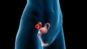
Spectral flow cytometry proves particularly valuable in research contexts where sample volumes are precious. In pediatric oncology, for example, researchers must extract maximum information from minimal cell numbers—a challenge where spectral systems excel by enabling comprehensive analysis from single samples.
Consider the reality faced by pediatric cancer researchers: By the time a child enters a research study, they have often undergone numerous procedures, making it difficult to obtain large bone marrow samples. The imperative becomes obtaining as many answers as possible, with certainty, from the smallest possible sample volume. Sometimes only enough material exists for 1 or 2 analysis tubes, demanding that every analytical tool maximize information extraction.
Spectral flow cytometry enables researchers to ask multiple questions simultaneously, facilitating faster, more precise decisions—sometimes from just a single, precious sample. This efficiency can make the difference between actionable insights and inconclusive results in critical research situations.
How spectral flow cytometry unlocks tumor heterogeneity
How is cancer complexity addressed?
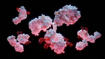
Cancer research has revealed that tumors are not uniform diseases but rather complex ecosystems composed of diverse cell populations that interact with immune systems in intricate ways. Understanding this heterogeneity has become essential for identifying new therapeutic targets and predicting treatment responses, challenges for which spectral flow cytometry is uniquely suited due to the following characteristics:
- Comprehensive immune profiling. Researchers can simultaneously analyze dozens of immune cell subsets, activation states, and checkpoint markers, providing unprecedented views of tumor microenvironments.10-13
- Immune evasion insights. By profiling both tumor and immune cells within a single analysis, investigators can uncover mechanisms by which tumors escape immune surveillance, directly informing next-generation immunotherapy development.
What does high-parameter analysis look like in practice?
Recent developments have demonstrated the power of extensive multiplexing in oncology research. A notable example involved developing a 50-color spectral flow cytometry panel that enabled comprehensive analysis of T cells, antigen-presenting cells, B cells, natural killer cells, and innate lymphoid cells within single experiments.10-12,14
This level of multiplexing revealed previously invisible details about immune cell differentiation and activation patterns, offering new avenues for therapeutic intervention that would have been impossible to identify using conventional approaches.10-12,14
How can autofluorescence be transformed from a challenge to an asset?
Autofluorescence has traditionally been considered a technical nuisance in flow cytometry, requiring compensation and subtraction to achieve clean results. However, spectral flow cytometry’s ability to distinguish and characterize autofluorescent signals has revealed their potential value as additional analytical parameters.
Changes in cellular autofluorescence can indicate disease progression or cellular transformation, providing researchers with supplementary markers for phenotyping that cost nothing additional but offer valuable biological insights. This transformation of a former limitation into an analytical advantage exemplifies how spectral approaches are expanding the information available from each sample.
Moving beyond manual analysis with AI and automation
As spectral flow cytometry panels grow in complexity, so too does the challenge of data interpretation. Recent advances go beyond basic automation. For example, artificial intelligence (AI)–driven tools are now being integrated to optimize panel design in real time, adapt gating strategies to novel sample types, and flag rare or unexpected cell populations for further investigation. These innovations are not only accelerating analysis but also helping to standardize results across users and sites, making high-dimensional cytometry more accessible to both new and experienced researchers.
How can barriers be reduced for new users?
These computational innovations prove particularly beneficial for laboratories with limited expertise in advanced data analysis. By automating complex processes, AI makes spectral flow cytometry more accessible and user friendly, further expanding its reach across diverse research communities.
The shift from subjective interpretation to objective, reproducible insights represents a fundamental improvement in how researchers can approach complex biological questions, regardless of their computational background.
How standardization can build trust
Ensuring that results are comparable across instruments and institutions is essential for research adoption. While industry-wide efforts, such as those led by the National Institute of Standards and Technology and Standardised Ontology Unique Labelling for Cytometry Annotation of Populations (SOULCAP), are establishing reference standards and shared nomenclature, many laboratories are also developing internal best practices for spectral panel validation and data quality control.2,15-18 These combined efforts are laying the groundwork for spectral flow cytometry to become a reliable tool for research discoveries.
Looking beyond cellular analysis
Technology providers play crucial roles in supporting researchers as they adopt spectral flow cytometry. Through close collaboration with customers, providers help streamline workflows, optimize assay design, and troubleshoot technical challenges.
This collaborative approach ensures that even laboratories new to spectral techniques can achieve high-quality, reproducible results. The partnership between technology developers and end users has proven essential for successful implementation, particularly as laboratories navigate the transition from conventional to spectral approaches while maintaining research productivity.
Flow cytometer operators can utilize different side scatters to separate distinct cell populations. By leveraging nontraditional side scatters—especially on red and infrared lasers—users can do label-free detection of cell populations, which enables unbiased, cost-effective identification of cells without external dyes or labels.
Dye chemistry is crucial in the workflow, with specialty dyes meticulously engineered to minimize spectral spillover and maximize brightness. These advancements are especially critical when designing complex, high-parameter panels for both conventional and spectral cytometry workflows.
By improving spectral fidelity, these innovations enable more flexible fluorochrome combinations, streamlining panel design and enhancing usability for researchers.
What emerging applications are on the horizon?
Spectral flow cytometry is poised to expand beyond traditional cellular analysis into new analytical territories, including:
- Noncellular entity analysis. Researchers are exploring applications for analyzing circulating tumor DNA, extracellular vesicles, and microbial populations using spectral techniques.
- Multimodal integration. Future developments may combine fluorescent and nonfluorescent measurements within single analytical workflows, enabling researchers to answer increasingly complex biological questions.
- Point-of-care applications. The potential for leveraging spectral flow cytometry in rapid diagnostic applications, such as pathogen identification or minimal residual disease monitoring, represents an exciting frontier for clinical implementation.
Conclusion
Spectral flow cytometry represents more than a technological advancement—it is a fundamental leap forward in the ability to profile complex biological systems with unprecedented precision and depth. The convergence of modular, accessible instruments with advances in AI, automation, and standardization is democratizing this powerful technology and enabling a new generation of discoveries across oncology, immunology, and beyond.
The transformation from intensity-based detection to comprehensive spectral analysis has opened doors previously closed to researchers, particularly those working with limited samples or in resource-constrained environments. As we’ve seen through examples in pediatric oncology and high-parameter immune profiling, this technology is not just improving existing workflows; it’s also enabling entirely new approaches to understanding biological complexity.
Looking ahead, the collaborative efforts of researchers, clinicians, and technology providers will be key to unlocking spectral flow cytometry’s full potential for both research and clinical care. The paradigm shift that began with capturing full emission spectra continues to evolve, promising even greater insights into the biological systems that define health and disease.
As adoption continues to grow and new applications emerge, spectral flow cytometry will further transform how we approach cellular analysis, moving us closer to a future where the complexity of biological systems can be fully understood and leveraged for human benefit.
Based on my 25 years of hands-on experience in flow cytometry, the impact that this technology can have in cancer research and especially in pediatric oncology is nothing short of life-changing. For me, this work is deeply personal. My family and I lost our beloved daughter, Lilly Anna, to cancer—a heartbreak that forever changed the way I see both life and science.
Her courage, light, and the painful journey we walked alongside her now live in every effort I make to support clinicians and researchers fighting for better outcomes for children like Lilly Anna around the world. Being in a position of influence within this field isn’t just professional—it’s sacred. I carry her name into every conversation, every innovation, every solution.
Beckman Coulter Life Sciences stood by my family and me during our darkest hours, not just as a company but as a community. Their support was deeply human and unwavering, and it affirmed for me that this work is not just about instruments or data. It’s about people, families, and hope.
About the author
Vasileios Toxavidis is senior solutions product manager at Beckman Coulter Life Sciences.
References
- Unlocking Insights: The Vital Role of Unmixing Algorithms in Spectral Flow Cytometry. Application note 2024-GBL-EN-106619-v2. Beckman Coulter Life Sciences; 2024. Accessed January 23, 2026. https://media.beckman.com/-/media/pdf-assets/application-notes/unlocking_insights_the_vital_role_of_unmixing_algorithms_in_spectral_flow_cytometry.pdf
- Spasic M, Ogayo ER, Parsons AM, et al. Spectral flow cytometry methods and pipelines for comprehensive immunoprofiling of human peripheral blood and bone marrow. Cancer Res Commun. 2024;4(3):895-910. doi:10.1158/2767-9764.CRC-23-0357
- Chattopadhyay PK, Gaylord B, Palmer A, et al. Brilliant violet fluorophores: a new class of ultrabright fluorescent compounds for immunofluorescence experiments. Cytometry A. 2012;81(6):456-466. doi:10.1002/cyto.a.22043
- Astakhova EA, Boeva AV, Demchuk AM, et al. Spectral flow cytometry: the current state and future of the technology. Int J Mol Sci. 2025;26(12):5911. doi:10.3390/ijms26125911
- Nolan JP, Condello D. Spectral flow cytometry. Curr Protoc Cytom. 2013;63(1):1.27.1-1.27.13. doi:10.1002/0471142956.cy0127s63
- Novo D, Grégori G, Rajwa B. Generalized unmixing model for multispectral flow cytometry utilizing nonsquare compensation matrices. Cytometry A. 2013;83(5):508-520. doi:10.1002/cyto.a.22272
- Ferrer-Font L, Mayer JU, Old S, et al. High-dimensional data analysis algorithms yield comparable results for mass cytometry and spectral flow cytometry data. Cytometry A. 2020;97(8):824-831. doi:10.1002/cyto.a.24016
- Beckman Coulter Life Sciences launches industry-first modular spectral flow cytometry solution. Beckman Coulter Life Sciences. News release. March 18, 2025.
- CytoFLEX Mosaic Spectral Detection Module: hardware and implementation. Technical specifications. Beckman Coulter Life Sciences. Accessed January 22, 2026. https://www.beckman.com
- Liu Y, Xu X, Liu D, et al. 30-color full spectrum flow cytometry panel for deep immunophenotyping of T cell subsets in murine tumor tissue. J Immunol Methods. 2023;516:113459. doi:10.1016/j.jim.2023.113459
- DeNiro G, Que K, Fujimoto T, et al. OMIP-105: a 30-color full-spectrum flow cytometry panel to characterize the immune cell landscape in spleen and tumor within a syngeneic MC-38 murine colon carcinoma model. Cytometry A. 2024;105(9):659-665. doi:10.1002/cyto.a.24886
- Longhini ALF, Fernández-Maestre I, Kennedy MC, et al. Development of a customizable mouse backbone spectral flow cytometry panel to delineate immune cell populations in normal and tumor tissues. Front Immunol. 2024;15:1374943. doi:10.3389/fimmu.2024.1374943
- Bonilla DL, Reinin G, Chua E. Full spectrum flow cytometry as a powerful technology for cancer immunotherapy research. Front Mol Biosci. 2021;7:612801. doi:10.3389/fmolb.2020.612801
- Konecny AJ, Mage PL, Tyznik AJ, et al. OMIP-102: 50-color phenotyping of the human immune system with in-depth assessment of T cells and dendritic cells. Cytometry A. 2024;105(6):430-436. doi:10.1002/cyto.a.24841
- NIST Flow Cytometry Standards Consortium. National Institute of Standards and Technology. Updated December 9, 2025. Accessed January 22, 2026. https://www.nist.gov/programs-projects/nist-flow-cytometry-standards-consortium
- FCSC Working Group 1—ERF-based instrument calibration and standardization. National Institute of Standards and Technology. Updated August 25, 2025. Accessed January 22, 2026. https://www.nist.gov/mml/bbd/fcsc-membership/fcsc-working-group
- SOULCAP—standardised ontology unique labelling for cytometry annotation of populations. SOULCAP. Accessed January 22, 2026. https://soulcap.org/
- Czechowska K, Bonilla DL, Cotty A, et al. Beyond the limits: how is spectral flow cytometry reshaping the clinical landscape and what is coming next? Cells. 2025;14(13):997. doi:10.3390/cells14130997
Articles in this issue
Advertisement
Related Content
Advertisement


Advertisement
Advertisement