|Articles|April 4, 2016
Applications of Microfluidics in Bioprocessing
Author(s)Randi Hernandez
Microchannels show potential benefits for inertial cell sorting and for introducing genetic material into cells.
Advertisement
Novel microfluidic technologies are advancing many aspects of biomanufacturing, including cell separation, sorting, and processing. Microfluidic perfusion devices, as described by Rajeev Ram, allow for automation of the fermentation process in the environment within a bioreactor. Ram
Aseptic cell sorting
In another application, microfluidic systems operated in parallel could replace membrane-based filters for microfiltration, according to research by Warkiani et al. (2). This could help eliminate bioprocessing problems related to membrane clogging and filter fouling, and has the potential to be incorporated into continuous processing for cell-separation systems with high yield. Cells are separated through hydrodynamic forces based on size in microchannels, a technique also known as inertial sorting. Through process modeling experiments, the investigators demonstrated that a microfluidic platform such as the one they examined could be capable of processing macroscopic-scale sample volumes. Compared with centrifugation, the acceleration of the cells traveling through the channels was modest, so the cells retained cellular homeostasis and were not under significant shear stress. The authors wrote, “Given the flexibility and relative ease of implementation of inertial microfluidics for passive sorting, we believe that this method of cell retention can be adopted easily in desktop perfusion bioreactors for continuous manufacturing of monoclonal antibodies” (2). After cells are sorted, microfluidic platforms also have applications for cell population heterogeneity and morphology analysis (3).
Introducing DNA into mammalian cells
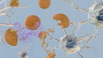
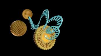
The fact that single cells can be isolated via microfluidics also has important implications for future transfection opportunities. Although foreign cellular material are most commonly incorporated into cells through chemical means (often by precipitation), by electroporation, or via microinjection, many of these techniques have the potential to adversely affect cell viability, and some of the methods are labor- and time-intensive. A highly efficient method of transfection, which has been tested in human cell types and has been highlighted in numerous peer-review articles, involves the mechanoporation of cells through microfluidic channels.
This “cell squeezing,” which was pioneered by MIT professors Klavs Jensen and Robert Langer and recently licensed to the company SQZ Biotech, offers an alternative method of delivering molecules into cells that may not be as disruptive as previous intracellular delivery methods. Larger proteins can get into cells more easily using mechanoporation, study authors have said, “without the need for extensive genetic engineering, protein engineering, biochemical modifications, and viral or synthetic packaging of protein” (4). SQZ Biotech’s CEO Armon Sharei reiterates that the company’s technique “has demonstrated the ability to deliver a broad range of materials, some of which are challenging to incorporate with current methods, to a variety of difficult-to-transfect cell types, including immune cells. By providing flexibility in application and obviating the need for exogenous materials or electrical fields, this method enables new avenues of disease research and treatment.” He adds that the only potential drawback of the method could be that the nature of the SQZ process limits it to ex-vivo cell-engineering applications.
Although repeated deformation of a cell seems like it could potentially be detrimental to a cell’s organelles (e.g., could cause damage to a cell’s endoplasmic reticulum, which could interfere with future protein glycosylation), Sharei assures this publication that “cellsqueeze” is engineered to minimize disruption of intracellular components. “Extensive work has been done internally to ensure cell health and functionality (both in vitro and in vivo) post-squeezing. Our results indicate that squeezing has far fewer functional side effects compared with other techniques, such as electroporation.”
References
1. R.J. Ram, "Tools for Continuous Bioprocess Development," BioPharm Int. 29 (1), pp. 18–25 (2016).
2. M.E. Warkiani et al., Sci. Rep. 5 (11018), doi: 10.1038/srep11018 (2015).
3. A. Grünberger, W. Wiechert, and D. Kohlheyer, Curr. Opin. Biotechnol. 29, pp. 15–23 (2014).
4. G.L. Szeto et al., Sci. Rep. 5 (10276), doi: 10.1038/srep10276 (2015).
Newsletter
Stay at the forefront of biopharmaceutical innovation—subscribe to BioPharm International for expert insights on drug development, manufacturing, compliance, and more.
Advertisement
Related Content
Advertisement


Advertisement
Advertisement
Trending on BioPharm International
1
First-in-Human Study Validates Safety of Next-Generation mRNA–LNP Platform
2
FDA Clears PharmaResearch IND for Nano-Based Cancer Drug PRD-101
3
How CDMO Alliances Can Provide End-to-End Service that Reduces Drug Development Time and Costs
4
High-Dose Nusinersen Slows Neurodegeneration in SMA Patients, Study Shows
5