
|Articles|May 4, 2022
- BioPharm International's BP Elements, May 2022
- Volume 1
- Issue 5
Standard BioTools Launches Hyperion+ Imaging System
Author(s)BioPharm International Editors
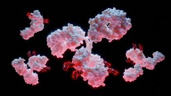
Standard BioTools’ new Hyperion+ Imaging System can process more samples and has lower limits of detection than their current product.
Advertisement
Standard BioTools, previously known as Fluidigm, announced the launch of the Hyperion+ Imaging System, a high-plex spatial imaging tool, in April 2022. Relative to the original Hyperion Imaging System, the Hyperion+ provides lower limits of detection, improved sample capacity, and quicker time to results.
According to an April 11 press release from the company, the system can process over 100 samples per week, approximately twice the speed of their current model. It also has a 1.6x lower limit of detection, enabling detection of dim markers, which can be important when analyzing the lower-expressing markers often used in translational studies.
The device allows for scanning of eight to over 40 targets in a single run and can easily be scaled to more than 100 targets. Additionally, by using an efficient, simple stain, image, and analyze workflow, it reduces expenses up to 10x relative to immunohistochemistry, which uses complicated cyclic immunofluorescence or digital profiling approaches, according to the release.
“Two critical challenges in realizing the full transformative potential of this remarkable technology are reducing the time to answer key biological questions and detecting important biomarkers that are expressed at low levels,” said Michael Egholm, CEO and president, Standard BioTools, in the press release. “The Hyperion+ Imaging System is designed to solve these challenges with faster time to results and a lower limit of detection than the current Hyperion Imaging System. These capabilities are key to quickly uncovering important spatial relationships with high-plex spatial imaging of 40-plus markers simultaneously at subcellular resolution.”
“My research group has been able to rapidly conduct exquisitely detailed phenotyping studies using the speed enhancements now available in the Hyperion+ Imaging System, which give us unprecedented insight into how cells within the tumor microenvironment function and interact with each other and the extracellular matrix,” said Bernd Bodenmiller, professor at the Department of Quantitative Biomedicine, University of Zurich, and at the Institute of Molecular Health Sciences, ETH Zurich, in the press release. “These insights are the foundation for innovating the next generation of cancer diagnostics and therapeutics and improving outcomes for patients with cancer.”
Source:
Articles in this issue
about 4 years ago
Shining a Spotlight on Nucleic Acid-Based Therapeuticsabout 4 years ago
Scaling Up Cell Therapies Capacityabout 4 years ago
MilliporeSigma’s ZooMAb Antibodies Earn ACT Label from My Green Lababout 4 years ago
Thermo Fisher Scientific Launches New GMP-manufactured Cas9 ProteinAdvertisement
Related Content
Advertisement

Advertisement
Advertisement