

A look at interchangeable biosimilars gaining FDA approval, the growth in global partnerships, and the rising biologics patent cliff.
A look at interchangeable biosimilars gaining FDA approval, the growth in global partnerships, and the rising biologics patent cliff.

The partnership expands patient access in MENA by localizing biosimilar manufacturing and distribution for gastro, neuro, and dermatology treatments.

The additional approval expands the label for Avtozma (tocilizumab-anoh) to now include the treatment of cytokine release syndrome, which aligns the therapy with all indications for which Actemra is approved in the US.
Under the licensing agreement, the proposed biosimilar candidate, PolyPB016, will be developed and manufactured by Polpharma Biologics and commercialized by Fresenius Kabi.

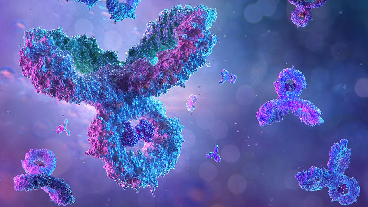
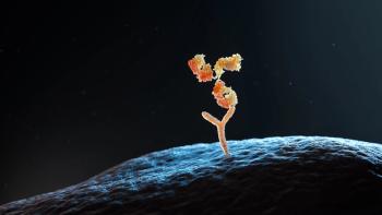
With more than 100 next-generation biologics set to lose patent exclusivity in the next 15 years, there is widespread agreement that developing biosimilars for these therapies will benefit society as a whole.

Biocon Biologics’ Yesintek demonstrated comparable safety and efficacy to the originator project, according to clinical data from a trial program.

As more originator biologics lose patent exclusivity in the next decade, a healthy biosimilars pipeline is necessary but requires market support.

The bio/pharma industry is evolving with intention, intelligence, and a growing sense of shared purpose.
Celltrion’s adalimumab-aaty is designated interchangeable as a high-concentration (100mg/mL) and citrate-free formulation of adalimumab.

Eculizumab-aagh (EPYSQLI) is now available in the US to treat patients living with difficult-to-treat rare diseases such as paroxysmal nocturnal hemoglobinuria, atypical hemolytic uremic syndrome, and generalized myasthenia gravis.
The EMA has issued a new proposal that aims to encourage biosimilar development and improve patient access to biosimilar therapies.
Process consistency and robustness, analytical excellence, and regulatory compliance are essential in the scale-up of biosimilars.
Omlyclo (omalizumab-igec) was approved to treat moderate to severe persistent asthma, chronic rhinosinusitis with nasal polyps, and certain food allergies.
The European Commission has approved Biocon Biologics’ YESINTEK, a biosimilar referencing Janssen Biotech’s Stelara (ustekinumab).
FDA’s Darby Kozak provided commentary on the anniversary of the Drug Price Competition and Patent Term Restoration Act of 1984.
Weighing development costs/resources and performance benefits is essential.
BioPharm International® spoke with Zara Bukhari, a PhD scholar at the University of the Pacific, Stockton, CA, and co-author of the peer-review research paper, Biosimilars Drug Development: Advances in Technologies from Molecule Design to Clinical Trials, published in the July/August 2024 issue, about process development for biosimilars.
The authors have reviewed applications of novel technologies in the major stages of biosimilars development: process development, pharmacology, toxicology, and clinical trials, with an emphasis on recent regulatory requirements.
STADA and Alvotech have launched Uzpruvo, the first approved biosimilar to Stelara (ustekinumab), in the European market, marking the second biosimilar brought to market by this partnership.
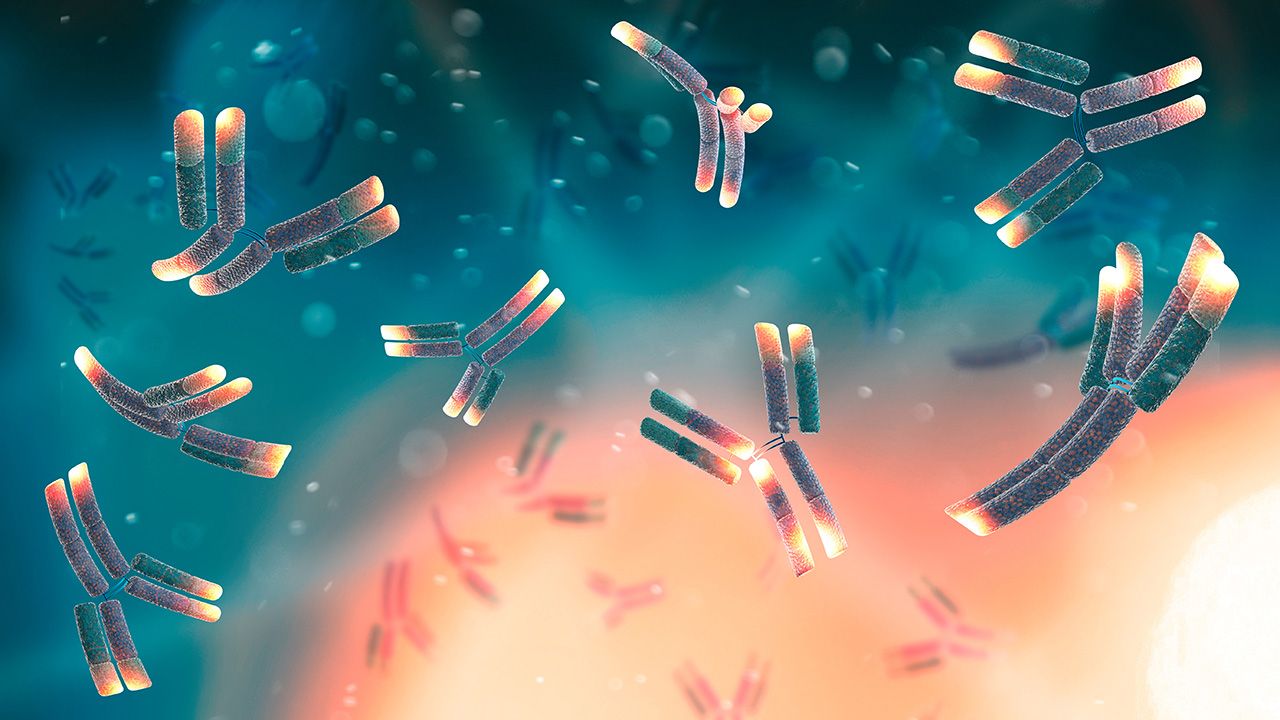
Biosimilar analytical assessments focus on demonstrating biosimilarity and interchangeability with the reference biologic.
Bkemv (eculizumab-aeeb) is the first interchangeable biosimilar to Soliris (eculizumab) to treat paroxysmal nocturnal hemoglobinuria and atypical hemolytic uremic syndrome.
Despite a growing number of biosimilar approvals, market uptake remains a challenge.
The European Commission has granted approval for Tyruko (natalizumab), a biosimilar developed by Polpharma Biologics for treating multiple sclerosis.
The EMA’s Committee for Medicinal Products for Human Use has given a positive opinion on Sandoz’ biosimilar trastuzumab for breast and gastric cancer.
Teva Pharmaceuticals and Alvotech will expand their partnership for the development and commercialization of biosimilar candidates.