|Articles|July 1, 2017
- BioPharm International-07-01-2017
- Volume 30
- Issue 7
Matching Tools to Biophysical Analysis Demands
Author(s)Guillaume Tremintin
Method choice is crucial to when seeking answers to biosimilar characterization questions.
Advertisement
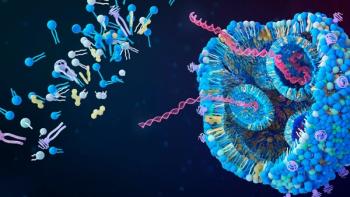
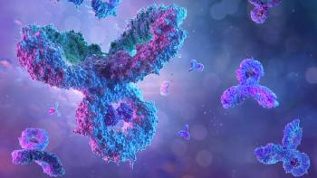
Monoclonal antibodies (mAbs) and related products continue to be the fastest growing class of human therapeutics. In 2016, for the first time, more than 30% of the total R&D pipeline was made up of biotherapeutics. Moreover, the sector recorded year-on-year growth from 2016 to 2015 of almost 16% (1).
As patents on therapeutic mAbs expire, the production of generic versions of these biopharmaceuticals (i.e., biosimilars) is a growing area of interest and importance. This article describes workflows and methodologies that can assist scientists as they develop biosimilars. Particular emphasis is placed on aligning the analytical question with the most appropriate tools available.
Analytical and regulatory challenges
A biosimilar-namely a copy of an approved biopharmaceutical-must be analytically proven to be highly similar to the original branded (innovator or reference) drug, with no differences in safety or efficacy (2,3). Making biosimilars, however, is fraught with challenges. mAbs and fusion proteins are glycosylated proteins (glycoproteins) produced by living cell systems such as mammalian cells, yeast strains, plant cells, or genetically modified animals. Each of these systems has its own glycosylation mechanisms, producing proteins with unique glycan patterns including different glycosylation sites and glycan composition (macro-heterogeneity) as well as different glycan linkages (micro-heterogeneity).
Although glycoproteins may have the same amino acid backbone, macro- and micro-heterogeneity create differing glycoforms of these glycoproteins (4,5).
The global regulatory landscape for biosimilar development is complex, with more than 30 national and international guidelines in place and a continuing debate around the role and relevance of preclinical and clinical in-vivo studies (6). What is consistent, however, is the early requirement for biophysical characterization of the biosimilar candidate in order to show that its glycosylation sites, glycan abundance, and structure are similar to the reference drug.
Mass spectrometry in biotherapeutics characterization
Mass spectrometry (MS) has long been seen as a powerful technique for elucidating the structure of proteins. Its applications include the identification of proteins and their post-translational modifications; the elucidation of protein complexes, their subunits, and functional interactions; as well as the differential protein expression in proteomics.
The two primary methods used for the ionization of protein in mass spectrometry are electrospray ionization (ESI) and matrix-assisted laser desorption ionization (MALDI). These ionization techniques are used in conjunction with mass analysers, such as quadrupole time of flight (QTOF).
In general, proteins are analyzed either in a top-down approach (in which they are analyzed intact) or a bottom-up approach (in which the glycoprotein is reduced enzymatically into small peptides and then analyzed). An intermediate middle-down approach, in which the mAb is cleaved under the hinge region and reduced before analysis, is often carried out. The subunit resulting from this preparation enables high resolution and high-dynamic range measurements.
Characterizing biosimilars
Work published around the time that the first biosimilar product was submitted for European regulatory approval (late in 2013) demonstrated how a combination of intact, middle-down, middle-up, and bottom-up ESI and MALDI MS techniques was able to characterize the amino acid sequence and major post-translational modifications in the marketed Cetuximab product (7). The approach described was suggested as a model for comparing mAbs and their biosimilar candidates.
The bottom-up approach to protein analysis in MS has been widely used for many years, whereby the protein is broken down into its peptides and separated using liquid chromatography (LC), before being identified through MS and matched to a database to find its most likely parent protein. Sample preparation for this workflow is long and involves multiple steps, and is known to potentially introduce artifacts or contaminants into the sample. More recently, top-down approaches or middle-down approaches have been employed, significantly reducing both sample preparation, and the potential of unwanted artifacts (8).
Each approach is suited to different applications and provides different data (Figure 1).
In practice
The traditional pillars of mAb characterization have been high-resolution ESI QTOF backed up by a peptide-mapping workflow. ESI QTOF gives a rapid analysis of the intact protein, but lacks sensitivity and the ability to resolve small mass differences. Adding peptide mapping to verify sequence information delivers huge dynamic range, but the workflow is time consuming and the digestion process is prone to creating artifacts in the sample. Using fluorescent labeling techniques, ESI QTOF is also routinely used to separate and quantify isometric forms of released glycans (9).
Recent work demonstrates how LC-free MALDI-TOF MS can give a quick overview at the released glycan level. (10) If little is known about the glycoprotein, or multiple glycosylation sites are present, analysis at the glycopeptide level can be useful. Through an LC-MALDI-TOF/TOF workflow, glycosylation sites and glycan composition can be determined (4). In addition, using LC-free MALDI peptide mass fingerprinting, more rapid glycoprotein identity testing can be performed. For fast profiling of quality attributes of a biosimilar and comparison with a reference mAb, analyzing the intact glycoprotein (top-down) or its subunits (middle-down) is the best option. These methods can provide a detailed profile with no or little sample handling required, thus reducing the potential for artifacts.
Analysis of a mAb subunit with an ultra-high resolution QTOF informs about the primary sequence and allows the rapid determination of the glycoprofile, which can then be compared with a reference database. Currently, top-down ESI MS of intact mAbs allows for only limited sequence coverage (approximately 30%), with incomplete sequencing of the complementarity determining regions, which are essential elements in antigen binding (11). More refined subunit middle-down experiments only cover 70-80% of the expected residues. In contrast, MALDI in-source decay (ISD) MS experiments have achieved close to 100% sequence coverage (12, 13).
In addition, studies with reference drugs cetuximab and natalizumab have shown that a combined approach involving middle-up mass measurement by LC-QTOF and middle-down protein sequencing by LC-MALDI ISD MS generated full sequence data (13).
Characterizing challenging modifications
Disulfides bonds may undergo scrambling under stress and may form derivatives such as thioethers or trisulfides (14). Trisulfide bonds are particularly challenging to deal with because any attempt to reduce the molecule destroys the information about the trisulfides. As such, it precludes using traditional inquiry methods such as subunit analysis or peptide mapping.
Antibody-drug conjugates are promising biotherapeutics for cancer treatment. High levels of trisulfides here, however, can interfere with the conjugation between the antibody and the drug, affecting efficacy (15). It is vital to verify early if a lead candidate is prone to trisulfide formation.
Recently, trisulfide structures have been identified in biopharmaceuticals using MALDI-MS (16). In this application, an automated method for identifying disulfide structures including scrambling byproducts was extended to include the detection of trisulfides (16). This identification was possible because trisulfide peptides show a specific fragmentation ion pattern that can be used for detection. In this work, the peptides in biopharmaceutical samples were separated using LC with MALDI-TOF/TOF to detect and identify the disulfide and trisulfide bonds. This approach was found to yield results with coefficient of variations of <20% (16).
Conclusion
Depending on the analytical questions being asked, different approaches can be taken to analysis and characterization. However, for the most common questions in the development of biosimilars--for which rapid profiling and comparison are required--top-down and middle-down approaches offer rapid fit-for-purpose solutions. MALDI is well proven in these applications, with full sequence validation possible. LC-free MALDI-MS can also accurately determine the molecular weight of the intact protein or domain at high speed and with ease. This workflow can also be used to determine glycan profiles from intact proteins or from released glycans. In addition, by performing LC-MALDI analysis at the glycopeptide level, glycan identification, and localization are also possible.
For intact glycoprotein, glycopeptide, and released glycan analysis, as well as middle-down analysis and/or in the characterization of chemical artifacts, MALDI is proving to be a vital analytical component. The technique is furthering the capabilities of MS in both innovator and biosimilar development and looks set to continue to do so in the future.
References
1. Pharma Intellegence/Informa,
2. F.M. Greer, 2012. “Biosimilars: The race to market continues...”, www.contractpharma.com accessed June 7, 2017
3. FDA, “
4. L. Zhang, S. Luo, and B. Zhang, mAbs, 8 (2) 205-215 (2016).
5. A. Varki and J.B. Lowe, “Biological Roles of Glycans” in Essentials of Glycobiology, A. Varki and R.D. Cummings, Eds. (Cold Spring Harbor, New York, NY, 2nd ed., 2009).
6. K. Chapman et al, mAbs, 8 (3) 427-435 (2016).
7. D. Ayoub et al, mAbs, 5 (5), 699-710 (2013).
8. J.A. Loo, I.S. Krull, and A. Rathore, LCGC North America 34 (6), 492-499 (2016).
9. T. Formorlo et al, State-of-the-Art and Emerging Technologies for Therapeutic Monoclonal Antibody Characterization, Volume 2. Biopharmaceutical Characterization: The NISTmAb Case Study; (American Chemical Society: Washington, DC, 2016).
10. N. de Haan et al. Anal. Chem. 87 (16) 8284-8291 (2015).
11. L. Fornelli, et al, Anal. Chem. 86 (6), 3005-3012 (2014).
12. J. Hardouin, Mass Spec Rev. 26 (5) 672-682 (2007).
13. A. Resemann, et al, mAbs, 8 (2) 318-330. (2016).
14. H. Liu and K. May, mAbs, 4 (1) 17-23 (2012).
15. K. Cumnock, et al, Bioconjugate Chemistry 24 (7), 1154-1160 (2013).
16. A. Resemann et al, “Towards the Fast and Increasingly Simplified Analysis of Trisulfide Formation in Biopharmaceutical Antibodies” (Presentation) CASSS Analytical Technologies, March 2016.
Article Details
BioPharm International
Volume 30, Number 7
July 2017
Pages: 44–47, 61
Citation
When referring to this article, please cite it as G. Tremintin, “Matching Tools to Biophysical Analysis Demands," BioPharm International 30 (7) 2017.
Articles in this issue
about 9 years ago
Laboratory Data Integrity Benefits from Electronic Systemsabout 9 years ago
Do You Listen to What Your Data are Telling You?about 9 years ago
Tale of Two Financial Trendsabout 9 years ago
Inspections: The Value of the Opening Presentationabout 9 years ago
Balancing Protocols for Leachablesabout 9 years ago
Biosafety Concerns of Single-Use Componentsabout 9 years ago
FDA Continues to Promote Quality Drug Productionabout 9 years ago
Contract Manufacturing Through the YearsAdvertisement
Related Content
Advertisement


Advertisement
Advertisement