
|Articles|August 1, 2021
- BioPharm International, August 2021 Issue
- Volume 34
- Issue 8
Using Picodroplet Technology to Optimize Bispecific Cell Lines
Author(s)Olivia Hughes
Growing demand for bispecific antibodies increases the need for automated and miniaturized high throughput screening capabilities.
Advertisement
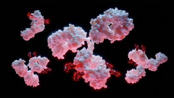
The field of biologics is rapidly evolving, offering new avenues for treating and preventing illnesses with unmet medical needs. The vast majority of therapeutic biologics have traditionally been monoclonal antibodies (mAbs), which account for nearly one-fifth of FDA’s new drug approvals each year (1). However, bispecific antibodies (BsAbs), which simultaneously target two different antigens, have emerged as the next novel wave of biologics (2).
These complex and highly potent molecules offer enhanced efficacy when compared to monovalent antibody therapeutics; however, BsAbs pose new challenges in development, such as low expression yields, inefficient heterodimer formation, and the provision of quality assurance data to regulatory agencies. Consequently, technologies that provide better predictive screening capabilities and higher throughputs than those traditionally used in cell-line development laboratories are required to meet the growing demand for new biotherapeutic molecules (3).
Bispecific antibodies—the next wave
Among antibody-based therapies, BsAbs have garnered considerable interest because of their additional functionalities. These functionalities include enhanced tumor killing by redirecting specific immune cells to the tumor cells, increased binding specificity by targeting two different cell-surface antigens, and enhanced therapeutic potency by inhibiting two biological pathways instead of one.
BsAbs are available in a variety of configurations, from antibody fragments without the fragment crystallizable (Fc) region to whole immunoglobulin Gs (IgGs) with the Fc region intact. Whole BsAbs are desirable candidates for novel therapeutics for cancer immunotherapy, inflammatory disorders, and other diseases. Typically, development begins with transfecting a host cell line with genes encoding the desired BsAb’s four peptide chains. However, BsAbs have a far more complex product profile than monospecific antibodies, containing a mixture of single peptide chains, monospecific half molecules, monospecific homodimers, and bispecific heterodimers, and this presents manufacturing challenges for BsAb cell-line development (4).
Specific challenges arise at various stages of development. Researchers must utilize sophisticated, expensive, and time-consuming analytical development techniques throughout, such as enzyme-linked immunosorbent assay (ELISA) and liquid chromatography–mass spectrometry to evaluate the structure, function, and quality of the products derived from each single-cell clone early in the process. Additionally, because the selection criteria for BsAbs are much stricter than those for monospecific antibodies, a much larger number of clones must be screened to identify cell lines with the highest quality and productivity.
Alongside these challenges, regulators in the pharmaceutical sector, such as FDA, require extensive proof of monoclonality for cell-line development. Manufacturers must provide visual evidence that each cell line is produced from a single parent cell to prevent costly delays in market authorization and clinical trial programs. Yet this critical regulatory requirement using traditional seeding techniques is labor intensive (3).
Enhancing cell line expression systems
In the highly competitive and economically driven biologics market, reducing the time to market is essential. Therefore, cell line development methods must strategically balance the need to deliver at speed without compromising the production of high-quality biologics (3). Cell culture processes can be designed to produce high titers of mAbs, and these methods can also apply to manufacture more complex molecules, such as BsAbs.
Stable mammalian cell lines are a well-established model system for the manufacture of recombinant proteins. Therapeutic BsAbs, particularly those with IgG-like properties, are created in mammalian cell lines such as Chinese hamster ovary (CHO) cells. CHO cells can produce fully humanized recombinant proteins with post-translational modifications, such as glycosylation, consistent with those found in human cells (3).
CHO cells provide versatility due to their ability to be cultivated in suspension, serum-free chemically defined media, and scaled up for long-term manufacturing needs. Transfection then results in a heterogeneous cellular population during cell line development, and cell lines are selected depending on desired characteristics. Accordingly, these cells expressing BsAbs should be able to produce the target proteins that are equivalent to the cells expressing the original therapeutic proteins.
Equally important is the use of appropriate multi-component vectors that optimize transcriptional regulation via elements such as promoters or enhancers and translation processes through initiation sequences, leader sequences, and polyadenylation signals. Additionally, the incorporation of appropriate selection marker genes to express the BsAb provides a vital detection flag that can be recognized quickly during the screening stages of cell line development (3).
Harnessing automated screening solutions
Novel, automated cell-line development technology can be leveraged to rapidly screen more cell types to select optimum cell lines capable of delivering high productivity and antibody quality throughout several generations. Picodroplet single cell analysis systems, based on the encapsulation of single cells into picolitre-volume aqueous droplets, have added a promising new dimension to cell line development and recombinant protein production. This high-throughput microfluidic technology improves the accuracy and efficiency of screening and culture development.
Miniaturized picodroplets provide a highly controlled microenvironment for studying individual cells, chemical and biochemical reactions, and cell–cell interactions while preserving cell viability, thus outperforming standard fluorescence-activated cell sorting (FACS). Picodroplets enable researchers to gain information on a single cell and the heterogeneity of a cell population that, otherwise, cannot be obtained using standard bulk assays (4).
Within a picodroplet, encapsulated cell-secreted molecules quickly accumulate to a concentration that can be measured consistently and quantitatively utilizing assays based on the Förster resonance energy transfer theory (FRET). Two fluorescent probes, the donor and acceptor, will attach to the target secreted molecule in a FRET experiment. The binding event brings the probes together, resulting in energy transfer from donor to acceptor, seen as a reduction in the donor’s fluorescence signal and a rise in the acceptor’s fluorescence signal. Picodroplets containing desired cells can then be fluorescently sorted and distributed into individual wells on a microtiter plate for downstream analysis (5). This procedure is carried out seamlessly within a picodroplet single-cell analysis device, resulting in a simplified, automated workflow (Figure 1).
A benefit of FRET is that it may be customized to detect two different BsAb specificities concurrently, allowing for fast identification of single cells generating properly constructed BsAbs (Figure 2). When used in conjunction with picodroplet technology, researchers can isolate single cells and trap all factors produced by those cells within a picodroplet that reduces diffusion and increases detection sensitivity. After just one to four hours of incubation, cell-secreted molecules reach a detectable concentration, permitting the analysis of the secretion profile and quality of the products from each cell clone within much shorter timelines (5). The ability to visualize single cells also allows for quicker and more accurate proof of monoclonality as required by regulatory agencies (3).
Due to the miniaturization of picodroplets, screening throughput is significantly greater than conventional techniques and more cost-efficient because of the miniaturized reagent requirements. By leveraging these enhanced single-cell analysis capabilities, researchers can now encapsulate and analyze hundreds of thousands to millions of cells in a single experimental run. Thus, providing a powerful tool to screen a vast cell population more efficiently and establish possible development clones in one day (5). As a result, fewer clones require downstream testing, allowing researchers to shorten overall project deadlines and conserve cell line development resources for BsAbs.
In addition to improved workflow performance and productivity, the new automated workflow frees scientists working in the cell culture and process development environments from tedious manual processes (5). Scientific teams can complete more projects in a given period while spending less time manually completing routine bench work. Compared to more traditional screening systems, automated picodroplet-based single-cell technologies are developed with integrated user-friendly software that require little training to apply, offering a low-risk, low-effort option to streamline workflows (3).
Picodroplet microfluidics
Picodroplet technologies have transformed single cell analysis in the discovery and development of novel therapeutics, enabling advancements in a number of applications, including BsAbs development. These complex molecules are forecast to be the next wave of biotherapeutics, providing greater patient benefits through their improved specificity and efficacy compared to monovalent antibody therapeutics (3). By using picodroplets to encapsulate single cells, these automated platforms not only enable more efficient processes, but offer significant cost savings. In doing so, picodroplet technologies can overcome many of the critical challenges in bispecific cell line development and ultimately increase productivity in biopharmaceutical discovery and development.
References
1. A. Mullard, “FDA Approves 100th Monoclonal Antibody Product,” www.nature.com, May 5, 2021.
2. S. Nie, et al., Antibody Therapeutics 3 (1) 18–62 (2020).
3. F. Saunders, International Biopharmaceutical Industry 2 (2) 42–45 (2019).
4. K. Matua, F. Rivello, and W.T.S. Huck, Adv. Biosys. 4, 1900188 (2020).
5. D. Josephides, et al., SLAS Technology 25 (2) 177–189 (2020).
About the author
Olivia Hughes, [email protected], is a senior marketing associate at Sphere Fluidics.
Article Details
BioPharm International
Vol. 34, No. 8
August 2021
Pages: 20–22
Citation
When referring to this article, please cite it as O. Hughes, “Using Picodroplet Technology to Optimize Bispecific Cell Lines,” BioPharm International 34 (8) 20–22 (2021).
Articles in this issue
almost 5 years ago
Subunit Vaccines and the Fight Against COVID-19almost 5 years ago
Cell Harvesting Sees Benefits from Automated Processesalmost 5 years ago
The Evolving Aseptic Processing Landscapealmost 5 years ago
Designing Flexible Fill/Finish Facilitiesalmost 5 years ago
Statistical Approaches for Determining Comparability of Biosimilarsalmost 5 years ago
Packaging for Stability Studies: to Outsource or Not?almost 5 years ago
FDA Prioritizes Vaccine Review and Process Improvementsalmost 5 years ago
FDA Inspections Back on Track?almost 5 years ago
GMPs for Emerging TherapiesAdvertisement
Related Content
Advertisement

Advertisement
Advertisement