- BioPharm International-11-01-2013
- Volume 26
- Issue 11
Metacomplex Formation and Binding Affinity of Multivalent Binding Partners
A study to investigate metacomplex formation in the hetero-association of a multivalent antigen, streptavidin (SA), and a bivalent antibody (Ab) using a composition-gradient multiangle light-scattering (CG-MALS) system consisting of a composition-gradient device, a MALS detector, and a UV/Vis absorption detector.
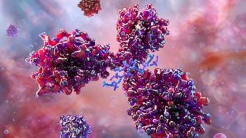
Standard analytical techniques for probing macromolecular interactions, such as surface plasmon resonance (SPR) or enzyme-linked immunosorbent assay (ELISA), require one binding partner to be immobilized on a surface to quantify the binding affinity. For simple 1:1 association and for some 1:n interactions, this physical tethering usually does not significantly impact the equilibrium dissociation constants (KD) that are measured. When both binding partners are multivalent, however, immobilizing one ligand can lead to erroneous estimates of the binding affinity by orders of magnitude due to avidity effects at the surface, mass transport limitations, and incorrect assumptions about the stoichiometry of the interaction. In contrast, composition-gradient multiangle light-scattering (CG-MALS) measures interactions entirely in solution, allowing all possible binding stoichiometries to occur and providing simultaneous quantification of self- and hetero-interactions, metacomplex formation, and other multivalent interactions (1). Figure 1 demonstrates the complex stoichiometries that result from the binding of an anti-streptavidin antibody (Ab) to the homotetramer streptavidin (SA).
Materials and Methods
CG-MALS experiments were performed with a composition-gradient system (Calypso II, Wyatt Technology Corporation) that prepared and delivered different compositions of protein and buffer to an inline UV/Vis detector (Model 2487, Waters Corporation) and a MALS detector (DAWN HELEOS, Wyatt Technology Corporation). Polycarbonate filter membranes with 0.1-μm pore size (Millipore) were installed on each fluid line for sample and buffer filtration.
Anti-streptavidin antibody was provided by Amgen. For CG-MALS experiments, streptavidin (Sigma-Aldrich) and antibody were diluted to a stock concentration of 10 μg/mL in a phosphate buffered saline (PBS, 25 mM NaH2PO4, 25 mM Na2HPO4, 50 mM NaCl, 200 ppm NaN3, pH 6.7) for a total of ~100 μg each protein per experiment. Each solution was filtered to 0.02 μm using Anotop syringe filters (Whatman GE) and loaded on the CG-MALS system (Calypso II, Wyatt Technology Corporation).
An automated CG-MALS method was run, consisting of single-component concentration gradients to quantify any self association and a dual-component crossover composition gradient to assess the hetero-association behavior (see Figure 2). For each composition, 0.8 mL of protein solution at the appropriate concentration was injected into the UV/Vis and MALS detectors. The flow was then stopped to allow the solution to come to equilibrium within the MALS flow cell. For single protein gradients, the flow was stopped for 120 s. The stop-flow time was increased to 500 s for the crossover gradient. A single experiment had an unattended run-time of ~3.5 h. Data collection and analysis of equilibrium association constants were performed using the manufacturer’s analysis software (CALYPSO software, Wyatt).
Results and discussion
The increased light-scattering signal in the hetero-association gradient region, corresponding to an increased weight-averaged molar mass, indicates that SA and Ab associate into complexes with a higher molecular weight than a simple 1:1 or 2:1 stoichiometric ratio (see Figure 3). Since neither protein was found to self-associate under these conditions, the higher-order stoichiometries must have resulted from the multivalent nature of the two binding partners.
In a crossover hetero-association gradient, the composition with the maximum light-scattering signal (weight-average molar mass, Mw) occurs at the overall stoichiometric ratio of the interaction. This ratio, combined with the magnitude of Mw, yield the absolute stoichiometry of the interaction. Figure 4 compares the measured Mw to three simple SA:Ab stoichiometries. For each simulation, the affinity per binding site was considered constant, KD = 0.2 nM. Although the measured data reach a maximum near the 1:1 molar ratio, the maximum measured Mw (~350 kDa; Figure 4, blue data points) is significantly larger than the maximum molecular weight for the 1:1 model (~200 kDa; Figure 4 dotted red line). A model considering two Ab bound per SA molecule approaches the correct maximum Mw (~330 kDa; Figure 4, solid purple line). This maximum, however, occurs at the wrong composition compared to the measured data, and this simulated model significantly underestimates the measured Mw for all compositions with excess SA.
Taken together, these data indicate the presence of higher order complexes with an overall stoichiometric ratio of 1:1, for example (SA)2(Ab)2, (SA)3(Ab)3, etc. This result seems surprising as SA’s four possible binding sites compared to the two on Ab suggest that complexes can occur with higher SA:Ab ratios as shown in Figure 1. The complex that saturates all expected binding sites would have an overall stoichiometric ratio of 2(SA):1(Ab). However, the crystal structure of SA shows a dimer of dimers with two-fold symmetry rather than a tetramer with four-fold symmetry (2), which could explain this propensity to form 1:1 metacomplexes rather than 2:1 metacomplexes. Interestingly, a similar 2:1 stoichiometric ratio for macromolecular interactions has also been observed with DNA aptamers raised against SA (3-5).
To characterize the SA-Ab interaction, two different models are considered: infinite self-association of [(SA)(Ab)] base units into n:n complexes, and a more refined analysis that includes n+1:n and n:n+1 complexes in addition to the n:n complexes.
First pass analysis: infinite self-association of 1:1 stoichiometries, [(SA)(Ab)]n
In the infinite self-association (ISA) model, SA and Ab bind with some affinity to form a 1:1 base unit [(SA)(Ab)]. This base unit then self-associates indefinitely, with each base unit adding to the growing chain with constant affinity. According to this model, Ab binds SA with affinity KD = 22 nM, and the [(SA)(Ab)] base units self-assemble with KD = 50 nM. An additional term accounting for two antibodies binding a single streptavidin molecule (SA)(Ab)2 was required to fully capture the change in light scattering as a function of composition. Under these conditions, there is no appreciable concentration of [(SA)(Ab)]n complexes with n > 3 (see Figure 5).
Refined analysis: inclusion of all (SA)i(Ab)j, for i,j < 3
Although providing a reasonable fit to the data, the ISA model implies that binding of a second partner to a divalent SA or Ab occurs with lower affinity than the first partner. While one might expect the affinities to be equal for these two symmetric proteins, this phenomenon could be explained by steric hindrance or allostery. A more complete analysis includes the n+1:n and n:n+1 complexes that should form in addition to the n:n complexes. Under this analysis (SA)(Ab)2, (SA)2(Ab), and (SA)3(Ab)2contribute significantly to the total light scattering (see Figure 6).
Other stoichiometries, including (SA)2(Ab)3 and (SA)2(Ab)4, drop out of the fit and do not contribute to the overall light-scattering signal. The lack of 2:4 and more extreme stoichiometries reflects the nature of SA to act as a dimer of dimers (i.e., contains only two effective binding sites for Ab). The apparent lack of a 2:3 complex is most likely a consequence of the sparse experimental data between the 1:2 and 1:1 composition ratios.
Our analysis yields KD values with a narrow range of 23 ± 4 nM per binding site. The constant affinity per binding site shows that the negative cooperativity predicted by the ISA model, disfavoring metacomplex formation, was an artefact of the initial analysis. The equivalence of all binding sites is reasonable since both molecules are symmetric and no allostery is expected. The residuals of this refined model are smaller and more random than those of the ISA model (see Figure 7), indicating a better description of the data.
Conclusion
Metacomplex formation by multivalent binding partners in solution is amenable to analysis by CG-MALS to obtain absolute stoichiometries and binding affinity. In this analysis, it was found that homotetrameric SA molecules present two binding sites for each bivalent Ab, leading to the formation of multiple stoichiometries in solution. By measuring the absolute molecular weight of the solution, CG-MALS determined the absolute stoichiometry of the complexes that were formed. Two different interpretations of the data have been presented: the ISA of n (SA)(Ab) base units into n:n complexes and a more refined model that added n+1:n and n:n+1 complexes to ISA. Under the latter model, (SA)(Ab)2, (SA)2(Ab), and (SA)3(Ab)2 complexes contribute significantly to the total light scattering, providing strong evidence for their presence.
The symmetry of each binding partner is reflected in the near constant affinity per binding site, KD = 23 ± 4 nM, confirming that there is no cooperativity favoring the formation of higher-order complexes or appreciable inhibition to their formation under these conditions. The number of complexes, ratio of SA to Ab, and degree of self-assembly could not be predicted a priori. By this analysis, CG-MALS proves uniquely suited to investigating complex systems of macromolecules in solution.
References
1. D. Some and S. Kenrick, “Characterization of Protein-Protein Interactions via Static and Dynamic Light Scattering,” in Protein Interactions, J. Cai, R.E. Wang, Eds. (InTech, Rijeka, 2012) pp. 401-426.
2. O. Livnah et al., Proc. Natl. Acad. Sci. 90 (11), 5076-5080 (1993).
3. V.J. Ruigrok et al., Chem. Biochem. 13 (6), 829-836 (2012).
4. Wyatt Technology Corporation, “Binding Affinity and Stoichiometry of a Multivalent Protein-aptamer Association,”
5. D. Some, Biophys. Rev. 5 (2) 147-158
(2013).
Articles in this issue
over 12 years ago
Supplier-Change Management for Drug-Product Manufacturersover 12 years ago
Report from Brazilover 12 years ago
Risk Aversion and Closed-System Processingover 12 years ago
The State of the Used-Equipment Marketover 12 years ago
Challenges in Managing the Cold Chainover 12 years ago
European Union Introduces GMPs for Excipientsover 12 years ago
USP Plans Standards on Good Distribution Practicesover 12 years ago
New Funding and Approval Pathways Prove Popularover 12 years ago
Disposable Applications in Demand by BiopharmaNewsletter
Stay at the forefront of biopharmaceutical innovation—subscribe to BioPharm International for expert insights on drug development, manufacturing, compliance, and more.


