
|Articles|June 1, 2017
- BioPharm International-06-01-2017
- Volume 30
- Issue 6
Particle Analysis Techniques Help Meet Regulatory Requirements
An increase in biologics raises awareness of particle generation and its role in negative patient outcomes.
Advertisement
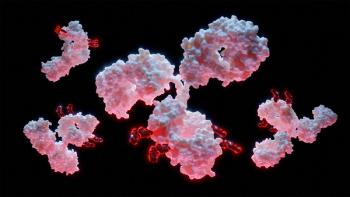
With the increase in biologic drug candidates, particularly monoclonal antibodies (mAbs) and high molecular peptides, the issue of particle generation has become more important due to their potential role in reduced drug activity and negative patient immune responses. These high molecular protein-based therapeutics are injected primarily either intravenously or subcutaneously, but also intravitreal. These therapeutics present challenges because they are less stable in liquid form than many small-molecule chemical entities. They can also have adverse reactions on contact with packaging materials and residue metals found in syringes or processing aids such as silicone oil. This instability leads to protein denaturing, loss in drug activity, and a potential to create long-term negative patient immune responses.
Regulatory authorities have responded to the issue of particle generation by enacting stronger regulations for particle detection and identification. FDA has engaged in an overhaul of their regulatory framework. And the US Pharmacopeial Convention has revised United States Pharmacopeia (USP) chapter <790>, and chapter <1790> has been adopted and provides guidance for 100% visual inspection of filled injectable units and possible particle characterization for GMP aseptic production (1, 2). The European Medicines Agency (EMA) has adopted similar regulations as well.
Authorities are now looking for a more expansive reaction to the discovery of visible particles during commercial production. They are focusing on particle characterization and root cause analysis of the type and source of particles generated in commercial batches.
Defining types and source of origin
Particle types can best be defined by the source of origin. For example, an environmental source such as cellulose fibers originating from disinfectant cloths, a human user, or metals emanating from filling equipment can lead to extrinsic particles. Intrinsic particles may result from inherent properties of the drug product, interactions of the drug product formulation components, or API combinations with primary packing materials or processing aids. They may also emanate from the active drug substance itself. Relevant particles range in size from subvisible in the nanometer range through to the micrometer range, crossing into visibility at approximately 100 micrometers.
Characterizing visible and subvisible particles
Until now, the standard procedure for quantification of subvisible particles in the micrometer range has been light obscuration using a liquid particle counter. This technique, which is a compendium method for routine testing used in batch release, determines the number and size of non-visible particles in the range of 1-100 µm. It has its limitations, however, especially because a description of the morphology of the particle and the chemical characteristics is not possible (Figure 1).
The standard procedure to determine visible particles is a manual visual check of the filled units and the subjective description of visible contaminants to demonstrate if any visible particles exist. Analytical labs are now establishing a range of novel analytical techniques to provide comprehensive particle characterization and identification. These techniques are primarily used to test and measure the limits of the compounding, mixing, and filling procedure design. They can also be applied to any issues arising in commercial batches by providing additional information to assist in determining root-cause analysis. The new methods include the
following:
- Micro-flow imaging (MFI). High-resolution images of subvisible particles between 1 μm and 70 μm are produced with MFI, which combines the possibilities of digital microscopy with modern microfluidics. Due to the morphology of the particles, modern application software makes it possible to determine particle characterization by number and size.
- Archimedes’ resonance measurement. With this technique, non-visible particles of a sample within the submicron range can be determined. This is achieved by transporting particles between 50 nm and 5 μm through a mechanically resonating microfluidic channel. The mass, dry mass, and size of the particle are then calculated. The difference in buoyancy of the particles allows for distinguishing extremely small subvisible particles such as silicone oil droplets or drug protein aggregates.
- Digital microscope coupled with Raman spectroscopy. Using a Raman spectrum, particles over a very wide range (1 μm-1000 μm) are classified by size, morphology, and chemical nature. This is accomplished by combining the automated static imaging capabilities of a high-resolution modern microscope with the chemical identification of individual particles using Raman spectroscopy.
- ETAC Proview (Bosch Packaging Technology)-digital visual inspection at laboratory scale. This computer-based automated inspection system is used for research and development purposes. ETAC Proview records and stores high-resolution photographs or film recordings of the filled units, making it possible to objectify the manual visual inspection appearance test. When combined with innovative application software, a precise evaluation of visible mobile and immobile particles within each unit can be achieved.
Conclusion
The increase in the number and nature of biologic drugs has led to stronger regulations from authorities that are expected to address the growing issue of particle generation. Manufacturers must enact these regulations and adapt accordingly. New tools are available for manufacturers to assist in testing both the size and nature of these particles, as well as any issues arising in commercial batches by providing additional information to assist in determining root cause analysis, and help to maintain the high standards of manufacturing quality.
References
1. USP, <790> “Visible Particulates in Injections,” USP 39–NF 34.
2. USP, <1790> “Visual Inspection of Injections,” USP 39–NF 34.
Article Details
BioPharm International
Volume 30, Number 6
June 2017
Pages: 30-31
Citation
When referring to this article, please cite it as M. Zerulla-Wernitz, "Particle Analysis Techniques Help Meet Regulatory Requirements," BioPharm International 30 (6) 2017.
Articles in this issue
about 9 years ago
Troubleshooting Lab Operations: Be Proactive, Not Reactiveabout 9 years ago
Unifying Continuous Biomanufacturing Operationsabout 9 years ago
N-Glycan Analysis of Biotherapeutic Proteinsabout 9 years ago
Efficient Manufacturing Critical for Accelerated Drug Developmentabout 9 years ago
Ensure Quality in a Contract Test Laboratoryabout 9 years ago
Storing and Shipping Frozen APIs in Single-Use Containersabout 9 years ago
Drug Spending Not Driving Rising Healthcare Costsabout 9 years ago
Managing Residual Impurities During Downstream Processingabout 9 years ago
A Novel Metric for Continuous Improvement During Stage ThreeAdvertisement
Related Content
Advertisement


Advertisement
Advertisement