|Articles|September 1, 2021
- BioPharm International, September 2021 Issue
- Volume 34
- Issue 9
In Vitro Pulls Ahead of In Vivo for Adventitious Agent Testing
Author(s)Meg Rivers
Scientific advances open the way to alternatives to animal testing and in-vivo assays.
Advertisement
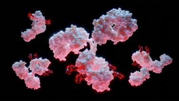
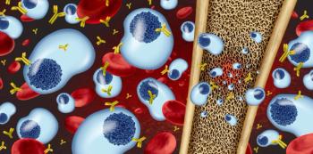
Adventitious agents—microorganisms introduced during the biomanufacturing process—present significant risks to the quality and safety of biologic drugs. Prevention, detection, and removal are key elements of risk mitigation practices.
“A source of risk for viral vaccines, biologicals, [and] cell and gene therapies are adventitious agents (AA)—microorganisms that may unintentionally be introduced into the manufacturing process,” explains Archie Lovatt, scientific operations director, SGS. “Exogenous forms of AA include bacteria, yeast, chlamydia, rickettsia, mycoplasma, prion, protozoa, and viruses. There are also endogenous forms of AA, which are process- or product-related impurities. These can include endogenous retroviruses or other viruses, replication-competent retroviruses, adenoviruses, or adeno-associated viruses, depending on the manufacturing process.”
Common sources of adventitious agents
“Within the biopharmaceutical industry, the main sources of adventitious agents are derived from the raw materials used during production, including the cell substrates, which are the starting point for the generation of biologics products,” says Alison Armstrong, global head of field technology management, MilliporeSigma. “Potential contaminants are also originating in manufacturing processes and cleaning procedures, equipment used, general production facilities used, and from direct and/or indirect human contact (i.e., personnel-related contaminations).”
The most challenging adventitious agents are new emerging viruses and viruses that can’t be assigned to a known virus family or are a distant variant of a known virus family, explains Horst Ruppach, executive director, scientific and portfolio, at Charles River. “Viruses that are not expected as a potential contaminant are especially difficult, such as the vesivirus contamination of [Chinese hamster ovary] CHO production cell lines or the porcine circovirus contamination found in rotavirus vaccines,” he says. “The latter was detected using high-throughput sequencing (HTS), mostly by chance. All typical testing assays applied during manufacturing failed to detect this virus.”
Key differences between in-vivo and in-vitro adventitious agent testing
Established in-vivo and in-vitro methods to test for adventitious agents have benefits and drawbacks.
“Both in-vivo and in-vitro adventitious agent tests are broad detection systems that can detect a wide range of viral contaminants,” said Armstrong. “The in-vivo assay is the classical method of virus detection used for many years to detect specific virus families, which allowed the detection of viruses not traditionally detected in cell culture-based systems. In-vivo assays use small animal models that meet regulatory requirements to provide an additional level of assurance of virus safety.”
Different guidelines for risk mitigation may require animal testing, says Pascale Beurdeley, chief scientific officer at PathoQuest. Animal testing is used for starting material evaluation (such as cell banks or virus seeds/vectors) and used only minimally for lot release. But one of the key differences between these two testing methods is timing.
“The turnaround time with animal testing is the longest: up to 42 days for the experimental phase of the in-vivo assays, two–six months for the complete study, as opposed to one week with molecular testing or four weeks for cell-based in-vitro testing,” Beurdeley continues. “Specificity and sensitivity of virus detection in in-vivo and cell-based in-vitroassays are usually unknown and currently, if an in-vivo or in-vitro cell-based result is positive (detection of adventitious contamination), it always requires additional investigation for virus identification using in-vitro or molecular assays.”
According to Ruppach, the breadth of detection and the sensitivity for specific viral contaminants can differ. The in-vitro assay typically has a higher breadth of detection and is more sensitive. A few viruses, however, are detected in the in-vivo assays at a higher sensitivity. For example, influenza contaminations can best be detected through inoculation in embryonated eggs.
“Different to the breadth of detection or the sensitivity, there are also some important practical aspects. The cell-based in-vitro assays typically perform better compared to assays using adult/suckling mice, guinea pigs, or embryonated eggs,” says Ruppach. “The administration of samples in vivo is tricky and sample matrix components can lead to animal death that could be misinterpreted as a viral contamination (false positive results). Both the in-vitro and the in-vivo assays require specialized laboratories but the requirements for in-vivo testing are even more challenging.”
Determining test suitability
Experts interviewed agree that in-vitro assays are usually the preferred method for detecting adventitious agents.
“In our industry experience, in-vivo methods have not proven to be effective in the detection of adventitious virus in products,” says Lorraine Borland, product manager, viral vaccine and gene therapy, Sartorius. “This may be due to lack of sensitivity of the method or indeed the reduced risk profile of products with the introduction of sterile single-use manufacturing systems, chemically defined medium, removal of animal components in the manufacturing chain, and increased process control.”
Scientific advances have opened the way to alternatives to using animals for testing. In some cases, replacing the use of animals with in-vitro testing are required.
“Given the range of technologies and testing strategies available today, it seems highly unlikely there are any instances where in-vivo testing would be considered more appropriate,” says Beurdeley. “Animal testing in general continues to fall out of favor in lieu of more novel molecular approaches like those that utilize the polymerase chain reaction, next generation sequencing (NGS), or combinations thereof.”
A 2014 study published in Vaccine by J. Gombold et al. found that some viruses can be better detected with in-vivo assays than with in-vitro cell-based assays—for example, the vesicular stomatitis virus and influenza virus. But in general, cell-based in-vitro assays have a higher potential to detect adventitious viruses (1).
“Interestingly, the in-vivo assay was included in the regulations for adventitious agent testing from the 1950s as a broad-spectrum detection system looking for viruses that were difficult to grow in culture and, therefore, could form contamination within a vaccine production stream without affecting the vaccine cultures themselves,” says Borland. “The hypothesis was that viruses, such as coxsackie virus, required the in-vivo test to be detected. For when they were isolated from clinical samples, it was seldom possible to grow them in cell culture unless a panel of cells were used. Isolation of the virus in suckling mice was, therefore, recommended. However, we can detect enteroviruses using in-vitro methods with a panel of susceptible cells and employing molecular methods.”
Strengths and weaknesses of in vivo vs. in vitro
Both in-vivo and in-vitro testing methods detect live contaminating viruses and have an amplification phase of contaminants, increasing the detectability of very low levels of contamination. Each testing method also has known strengths and weaknesses.
“For in-vitro assays, the use of selected cell lines is permissive to a wide range of viruses using several end-point test systems (i.e., cytopathic effect, haemagglutination inhibitionor haemadsorption, or immunofluorescent assays [IFA]),” says Armstrong.
Although both assay types detect infectious viruses, neither are usually specific, says Ruppach. Ruppach adds that a viral contaminant isn’t automatically identified in a positive result. The exception is when applying specific read-out methods like ELISA or polymerase chain reaction (PCR), which are designed for specific viruses.
“I don’t see much strength with the in-vivo approaches except if a specific contaminant is assumed and known to be best detectable through animals,” says Ruppach.
Current adventitious agent tests can detect a broad range of viruses at a low limit of detection of about 1 TCID50, Armstrong explains. Because these test methods have a limit for the sensitivity they can detect, any contaminants below that level are challenging to detect. Armstrong further adds that known limitations for cell-culture-based systems include the fact that not all virus agents grow well in cell-culture-based systems. Therefore, according to Armstrong, there is a need for alternative methods in adventitious agent testing—for example, molecular-based technologies including degenerate PCR methods, advanced molecular technologies such as NGS/HTS or IFA systems, which allow detection of a wider range of known and emerging adventitious agents.
“Latent or silent virus infections can be also difficult to detect and require special testing regimes” says Armstrong. “Cell culture systems are particularly susceptible to matrix-related toxicity, as well as to viral infection-related cytopathic effects.”
Knowing the benefits and shortcomings of adventitious agent testing, what is next for the industry? NGS or HTS technologies are considered suitable supplements or even replacements of in-vivo and in-vitro assays, says Ruppach. In a given sample, these technologies detect nucleic acids—including virus-specific nucleic acids—and analyze the nucleotide sequence. Regulators have recognized the impact of state-of-the-art technologies and have provided guidance on the use of such technologies (WHO TR978, recent EP chapters 2.6.16, 5.2.3, and 5.2.14).
“The major benefit is that these methods can detect and identify all viruses (even unknown viruses) at a sensitivity compared to PCR or even beyond. In fact, there are ongoing discussions that NGS/HTS may ultimately replace the in-vivo methods for both 3Rs considerations, and the limitations outlined above,” Ruppach says. “The major difference between NGS/HTS to the in-vitro and in-vivo methods is that NGS/HTS methods detect both infectious and non-infectious viruses. Non-infectious viruses or related nucleic acids could be a left over from a treatment, and—while not typically a risk—they can lead to a ‘false’ positive result with an NGS/HTS approach. However, while NGS/HTS methods have been developed to differentiate infectious from non-infectious viruses, which could make these technologies universal for viral contamination detection moving forward, there are still misconceptions about their effectiveness [according to the literature (2)].”
Beurdeley adds, “Both tests [in-vivo and in-vitro cell-based assays] were initially assessed as broad range virus detection assays. This approach has been challenged with the advent of truly broad molecular test methods, such as NGS, which does not require prior knowledge or suspicion of the adventitious agent. Moreover, our alternative NGS approaches can distinguish replicative—thus infectious—from non-replicative viruses. As such, NGS can detect previously undetected or undetectable replicative virus contaminants [citing an article in the Journal of Virology (3)].”
Only time will tell what technologies will emerge most effective for detecting adventitious agents and other contaminants.
References
- J. Gombold, et. al., Vaccine, 32(24) 2916–2926 (May 19, 2014).
- A. Brussel, “The Emergence of Next Gen Sequencing for Analyzing Biologics,” Eureka, Charles River blog, July 7, 2021.
- J. Victoria, et al., Journal of Virology, 84(12) 6033–40 (June 2010).
About the author
Meg Rivers is a senior editor at BioPharm International.
Article details
BioPharm International
Vol. 34, No. 9
September 2021
Pages: 44–47
Citation
When referring to this article, please cite it as M. Rivers, “In Vitro Pulls Ahead of In Vivo for Adventitious Agent Testing,” BioPharm International 34 (9) 44–47 (2021).
Articles in this issue
almost 5 years ago
Setting a Clear Strategy for Primary Packagingalmost 5 years ago
Bioprocessing Innovations Pose New Challenges for Fermentationalmost 5 years ago
Process Chromatography Makes Steady Progressalmost 5 years ago
Reevaluating Cell and Gene Therapy Developmentalmost 5 years ago
Using Digital Twins to Model Process Chromatographyalmost 5 years ago
Opportunities for Digital Twinsalmost 5 years ago
How Pure is Pure? Understanding Reagent Purity Gradesalmost 5 years ago
The Impact of Insufficient Oversightalmost 5 years ago
Trends Affecting Biopharmaceutical ManufacturingAdvertisement
Related Content
Advertisement


Advertisement
Advertisement