


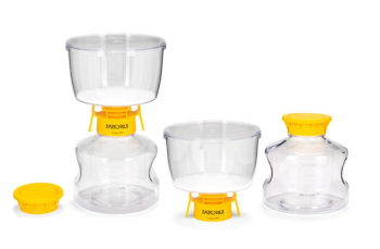
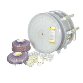
Sartorius’ Sartolab RF|BT 150–1000 vacuum filtration units provide fast, high-capacity filtration.
Sartorius’ Sartolab RF|BT 150–1000 vacuum filtration units provide fast, high-capacity filtration.
The MicroEDS BioWaste Treatment System can process between 150 to 500 L of waster per day.
The 3M Harvest RC clarifier condenses three process steps into a single-stage purification of recombinant protein therapeutics.
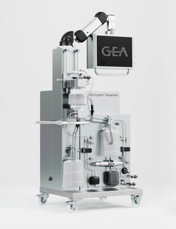
The GEA kytero combines the high performance of larger GEA stainless-steel pharmaceutical centrifuges with the benefits offered by disposable components.
STERIS Life Sciences’ VestaSyde SQ 64 st Ready-to-Use Disinfectant Wipes are part of a family of products designed to disinfect cleanroom services.
SKAN’s new Cellana isolator is designed for GMP manufacturing of ATMPs.
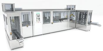
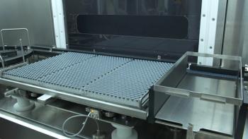
Syntegon's Syringe Inspection Line utilizes an AI vision system for the inspection of syringe flanges, stoppers, and cylinders.
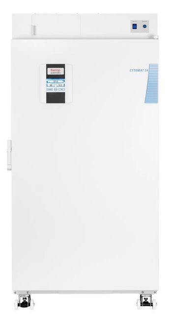
The Cytomat 24 automated incubator system is designed for large-capacity microplate processing.
SP Scientific's SALS30 and RxR-36 are designed for fill/finish processing applications like freeze dryers.
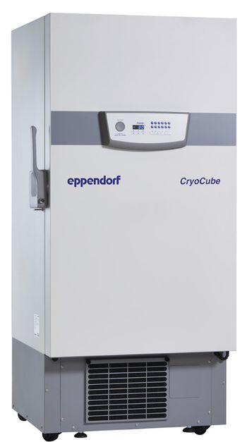
Eppendorf's CryoCubeF440 freezer has been redesigned to promote sustainability through various green features.
SP Scientific Products’ SP Hull LyoStar 4.0 R&D freeze dryer supports rapid freeze-dry cycle development, optimization, and process scale-up.
CalAmp's new sensor for cold-chain applications provides visibility for shipping vaccines.
Freezers, shipping containers, and tracking systems aid distribution of COVID-19 vaccines.
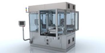
Stevanato Group’s Vision Robot Unit uses AI-based machine learning capabilities for particle and cosmetic inspection of biopharmaceutical drug products.
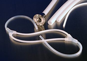
AdvantaPure’s GORE STA-PURE Pump Tubing systems can now overmold into any manifold tubing assembly as its pump element.
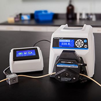
The new Masterflex Flow Controller from Cole-Parmer measures, controls, and logs fluid path information after receiving input from a flow sensor and then modifies the speed of the pump to support a set flow rate or dispense volume.
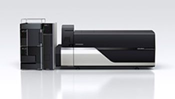
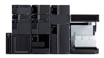
The LCMS-8060NX from Shimadzu is a triple quadrupole mass spectrometer that works to enhance method development and routine analysis.
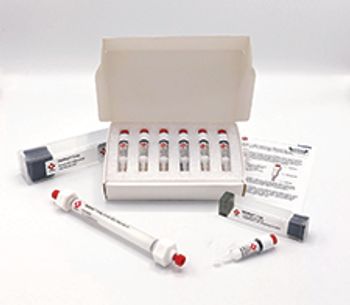
The company now offers its CONFIDENCE virus clearance services to support validation of viral clearance processes.
Honeywell Forge Cybersecurity Suite’s improved industrial-grade remote access and added protection and risk monitoring aids remote operations.
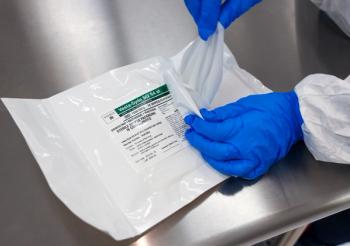
The VIA Capsule from Cytiva is a liquid nitrogen-free cryogenic shipment system designed to transport cell therapies.
Remote, machine health monitoring services reduce the need for on-site visits.
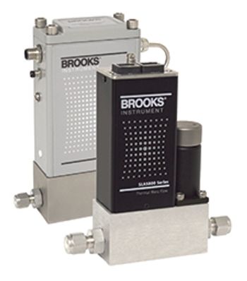
The SLA5800 Series Biotech and the SLAMf Series Biotech are two new mass flow controller (MFC) models from Brooks Instrument designed specifically for improved gas flow control in biotechnology applications.
Tosoh Bioscience introduced the SkillPak 1 mL and 5 mL pre-packed columns for fast method development and resin screening of monoclonal antibodies, antibody constructs, oligonucleotides, proteins, and viruses.
The Nexera UC Prep, a preparative supercritical fluid chromatography (SFC) system from Shimadzu, works to provide maximum use of lab resources through flexible system configurations in a compact design that requires low installation space.
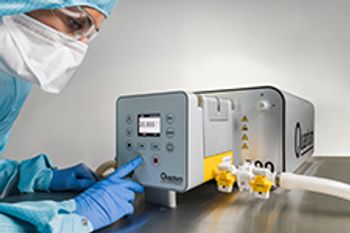
ReNu SU (single-use) Technology cartridge assemblies from Watson-Marlow Fluid Technology Group are designed for the development and production of personalized medicines.