
|Articles|November 1, 2015
- BioPharm International-11-01-2015
- Volume 28
- Issue 11
Implications of Cell Culture Conditions on Protein Glycosylation
Author(s)Dr. Richard Easton, Michiel E. Ultee, PhD
The authors present a review of the techniques commonly used for glycosylation analysis.
Advertisement
This article reviews the implications of cell-culture conditions on biologic product quality, focusing on glycosylation and analytical techniques for its accurate assessment. Glycosylation can potentially affect a protein’s half-life, immunogenicity, binding activity, and stability. It is a complex process that consists of the attachment of carbohydrate moieties, with possible attachment sites via asparagine (N-linkage) or serine/threonine (O-linkage) amino acids in protein structures. In mammalian cell culture processes, the use of different species can potentially produce significant differences in the types of glycosylation that can occur. These differences in glycosylation can have significant effects on the quality of the therapeutic protein produced, as can the choice of cell clone, the basal and feed media used, and the cell-culture conditions.
The choice of host cell and the bioreactor conditions used in bioproduction of proteins significantly affects protein product quality. This is due both to the structural complexity of proteins themselves and also to species-specific post-translational modifications that may occur during the cell-culture process, glycosylation being of particular importance.
Protein therapeutics and cell-culture effects on protein quality
Biopharmaceutical drugs are proteins with polymeric structures, built up in a series of structural levels starting from the amino-acid sequence, referred to as primary structure, through folding of the amino acid chains into local (secondary) and longer-range (tertiary) three-dimensional conformations. Multi-chain proteins, such as IgG antibodies, additionally have a quaternary structure resulting from structural associations between the subunits.
The choice of host-cell line for recombinant protein production depends first on the protein’s molecular properties. Certain bacteria can be used for production of the simplest proteins, those that are composed only of amino-acid polymers, with no post-translational modifications (PTMs) such as glycosylation, because most bacterial strains are incapable of glycosylation. Production is fast using simple media; however, purification can be challenging. Rapid production of proteins with primitive glycosylation can be achieved using yeast. Insect cells, generally used with a baculovirus vector in transient fashion, are used mostly for R&D and niche products. Mammalian cells are used for the production of complex proteins such as antibodies and enzymes, requiring full PTMs, including the production of complex carbohydrates.
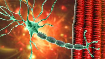
Figure 1: Graphic showing IgG antibody structure, sites for glycosylation, and potential variability.Proteins are delicate molecules compared with small-molecule drugs and present multiple stability challenges. A typical glycoprotein such as an IgG antibody has many sites of variability within its structure, which comprises four chains with a total molecular weight of 150,000 Da. Additionally, there are several post-translational modifications of the protein chain that can occur, such as oxidation and deamidation of specific amino acids. Each heavy chain also includes a site where glycosylation takes place (Figure 1).
Why is glycosylation important?
Many complex proteins such as antibodies and enzymes are glycoproteins, containing from 2–30% carbohydrate. Glycosylation is a complex process, with the carbohydrate attached to the protein either via the amino acids asparagine (N-linked) or serine/threonine (O-linked). Multiple sugar types are possible, each with multiple attachment sites, and variation of the mammalian cells used in production can lead to subtle glycosylation differences (1).
The choice of cell clone affects product quality. Each clone has slightly different abilities for glycosylation and other PTMs, and viabilities vary, resulting in differences in released intracellular degradative enzymes into the culture. Therefore, it may be necessary to select a cell clone that is not the highest producer to achieve the desired protein quality.
The extent of glycosylation can also vary depending on the basal and feed media, even with a single clone producing a single monoclonal antibody, or single basal media with varied feeds.
Effects of cell-culture conditions
Cell-culture parameters that affect the type and extent of glycosylation include pH and CO2 levels; the amount of dissolved oxygen (dO2); the temperature; the levels and types of nutrients; the presence and types of glycan precursors; cell viability, as dying cells release degradative enzymes; and the level of process control.
Bioreactors offer much greater control of pH and dissolved gasses than shake flasks do, and hence, better process control, but shake flasks are more economical and readily allow larger numbers or arrays. Shake-flasks are, therefore, commonly employed for early scouting studies on media and feed conditions. Optimum cell-culture development is achieved by working with bioreactors to enhance growth and productivity via selection of basal media and culture feeds and the timing for these feeds; optimizing bioreactor oxygenation conditions, such as CO2 levels, pH, agitation, temperature, seeding densities, and split ratios; extending the production phase of cell culture through the use of temperature shift; and minimizing accumulation of growth-inhibiting metabolites such as ammonia.
More recently, the availability of miniaturized bioreactors has provided an effective and efficient way of conducting multi-parameter studies. Thus, ‘Big-Data’ approaches allowing design-of-experiment (DoE) studies, in which multiple inter-reacting bioreactor conditions are evaluated simultaneously, are made possible. Such studies require the employment of automated, large-number bioreactor arrays to make the process feasible (2).
Special cell-culture considerations for biosimilars
Biosimilars are “generic” versions of protein pharmaceuticals that must be highly similar to the innovator drug in order to be classified as such. Regulatory guidelines require extensive analytical testing side by side with the innovator drug, including full glycosylation profiles (3). Similarity to the innovator drug is paramount; this must begin from clone selection and proceed throughout process development. Therefore, rather then only selecting for clones with the highest titer, as is usually the case for innovator drugs, selection is first for biosimilarity, which may mean that some of the highest producing clones are not selected.
Techniques for glycosylation analysis
N-glycosylation occurs when the carbohydrate is attached to asparagine in the consensus sequence asparagine-X-serine/threonine, where X is any amino acid except proline. As a nascent protein is being synthesized in the endoplasmic reticulum within the cell, an en bloc transfer of a pre-formed, lipid-anchored conserved glycan occurs. As the synthesized protein makes its way through the Golgi apparatus, the conserved N-linked glyan is ‘processed’ by enzymes (glycosidases and glycosyltransferases). It is the presence of these processing enzymes, their relative levels, and the accessibility of the glycosylation site on the protein to these enzymes that determine the final glycosylation of a protein.
O-glycosylation occurs on the amino acids serine and threonine, but not in accordance with a linear consensus sequence. There are some rules governing the process-for example, there are often nearby proline amino acids within the regions of glycosylation, and also quite often, tandem repeats of serine and theonine.
Analysis of N-glycans
Analysis of N-glycans is the most active area of research. The techniques that apply to analysis of N-glycosylation also apply to O-glycosylation.
The most abundant mammalian N-glycan structure is the complex type where a number of N-acetylglucosamine structures are appended to the molecule and extended with galactose, fucose, and sialic acid residues to give between two and four antennal structures. The exact structure reflects the nature of the enzymes present in the cell type used in the expression process as well as the precise environment in which the cells are located.
International Conference on Harmonisation (ICH) Quality Guidelines Q6B describe the type of analysis that should be performed in relation to these structures to understand the quantities of the different types of monosaccharides present, the nature of the glycans in terms of their antennary profile, and linkage of monosaccharides in each structure but also the structure of the glycans at the different glycosylation sites on the protein backbone (4).
Analytical procedure: The details
The analytical procedure of glycosylation involves a number of physical and enzymatic steps to release the glycans and then separate them from the peptides and any O-glycopeptides present in the mixture. O-glycans can be released chemically from these O-glycopeptides and purified separately from the remaining peptide chains.
A permethylation derivatization procedure is then performed on the separated N-glycans and O-glycans, enabling them to be analyzed using matrix-assisted laser desorption ionization–mass spectrometry (MALDI–MS) analysis.
It is also possible to analyze the samples chromatographically without having to employ the permethylation procedure. For example, a high pH anion-exchange chromatography procedure with pulsed amperometric detection (HPAEC–PAD) can be used for the analysis of N-glycans. An example of this can be seen in the N-glycans released from bovine fetuin, as shown in Figure 2. This method gave consistent data across three preparations, a series of clustered peaks representing di-, tri-, and tetra-sialylated glycans (i.e., two, three, and four sialic acid groups on the N-glycans within each cluster). The analysis gives some structural information in terms of an idea of what has been produced but little information in terms of the precise nature of what the glycans are. Techniques like this are, therefore, useful for comparative work but do not give much structural information for characterization of the molecules.
Figure 2: Stacked high pH anion-exchange chromatography procedure with pulsed amperometric detection (HPAEC-PAD) chromatograms of glycans released from three preparations of bovine fetuin.In an alternative approach, a fluorescence tag-2-aminobenzamide-can be used to specifically label the released N-glycans and these labeled N-glycans can then be chromatographically separated. The chromatographic eluent is then analyzed by mass spectrometry (MS) to identify the masses of individual structures. This is a useful technique because the chromatographic profile not only gives a batch-to-batch comparison but also provides information on the masses of the glycans in each peak. The technique is so sensitive that it is possible to resolve and identify the two different forms of the so-called G1F structure, the monogalactosylated biantennary glycan found on antibodies. The two components have a galactose structure on either arm of the biantennary structure and these can be separated out. Further use of mass spectrometry, however, is necessary to fully characterize what these glycans are.
Characterization using mass spectrometry
The aforementioned permethylation procedure allows the separation of glycans with similar masses in the non-derivatized state. For example, two glycans-with identical structures apart from one containing a single N-aetylneuraminic acid (sialic acid) residue and the other containing two fucose residues-differ in molecular weight by only 1 Da in the native state but differ by 13 Da when derivatized. Matrix-assisted laser desorption ionization–mass spectrometry (MALDI–MS) analysis gives a population profile of the glycans that are present. Using the peaks (masses) of the spectrometric analysis, in combination with the knowledge of the N-glycan, the core structure, and the biosynthetic pathways that are present, it is possible to arrive at glycan monosaccharide compositions. However, this leaves the arrangement of the glycans within the structure to be determined.
Next, electrospray–mass spectrometry (ES–MS) can be used to fragment the permethylated glycans. This division action is useful, as the fragmentation pathways are well defined, well understood, and produce specific masses depending on where the fragmentation takes place. This process depends on the exact nature of the structure of the N-glycans so these fragment masses allow the determination of the antennary structures present on the glycans.
A third method, gas chromatography–mass spectrometry (GC–MS), can be performed to determine how the monosaccharides are linked to one another within the glycan structure. These structural variations can have a large effect on the drug’s efficacy and properties.
By chemically modifying the permethylated glycans and attaching particular “reporter groups” wherever each monosaccharide is linked one to another, derivatives are produced that provide key structural information when analyzed by mass spectrometry. Using an electron-impact mass spectrometer, the fragmentation patterns show characteristic fingerprints for the different linkages. Furthermore, the GC apparatus on the front end of the mass spectrometer can identify each of the different linked structures with their different reporter groups, because these have different elution times from the GC column. The combination of GC retention time and mass spectral fingerprint fragmentation pattern identifies the different monosaccharide linkages that are in the glycan population.
One final step in characterization is to determine the stereospecificity of any linkages within structures where data indicate that potentially immunogenic epitopes such as GalαGal (a known immunogenic epitope) might be present. Treatment of the glycan with a specific exoglycosidase enzyme and analysis by MS pre- and post-exposure can confirm the presence of an alpha-linked galactose by revealing a mass shift upon incubation.
Conclusion
Proteins are inherently complex and much larger in size than small-molecule drugs, and multiple types of PTMs, including glycosylation, are a feature of most protein therapeutics. Their occurrence depends on the molecular nature of the protein; the selection of cell type and individual clone; and cell-culture and bioreactor conditions. Extensive analytical assays are needed to characterize a protein therapeutic. Biosimilars require even more analytical testing than do innovator drugs because of the need to prove biosimilarity to the given innovator molecule.
Mass spectrometric analysis provides structural information on the composition, antennal structures, and linkages present in N-glycan molecules and the use of chromatography provides a unique glycan profile due to the precise structure and associated interactions of the glycans with the column matrix. This profile can be used as a reference against which other batches can be compared. In addition, the use of LC/ES-MS of proteolytic digests allows sites of glycosylation to be isolated and identified. These sites can then be analyzed using a number of techniques to determine the nature of the glycans at each site and the specific populations of the glycans on the biopharmaceutical molecule.
References
1. D. Ghaderi et al., Biotech Gen. Eng. Rev. 28, pp. 147–176 (2012).
2. S.D. Jones et al., Am. Pharma. Rev. 18 (1), pp. 44–48 (2015).
3. FDA, Guidance for Industry, Scientific Considerations in Demonstrating Biosimilarity to a Reference Product (Rockville, MD, Apr. 2015).
4. FDA, Guidance for Industry, Quality Considerations in Demonstrating Biosimilarity of a Therapeutic Protein to a Reference Product (Rockville, MD, Apr. 2015).
5. ICH, Q6B, Specifications: Test Procedures and Acceptance Criteria for Biotechnological/Biological Products, Step 5 version (Sept. 1999).
About the Authors
Michiel E. Ultee, PhD, is principal at Ulteemit BioConsulting, LLC, and Richard Easton, PhD, is team leader, carbohydrate analysis, SGS M-Scan Ltd.
Article DetailsBioPharm International
Vol. 28, No. 11
Pages: 20–25
Citation: When referring to this article, please cite it as M. Ultee and R. Easton, "Implications of Cell Culture Conditions on Protein Glycosylation," BioPharm International28 (11) 2015.
Articles in this issue
over 10 years ago
Selecting the Right Viral Clearance Technologyover 10 years ago
Best Practices for Sterility Assurance in Fill/Finish Operationsover 10 years ago
CMOs Continue to Improve Overall Biomanufacturing Performanceover 10 years ago
FDA Overhauls Inspection Operationsover 10 years ago
Good Documentation Practice: Saving Data for the Long Termover 10 years ago
Next Steps in Outsourcing Relationshipsover 10 years ago
Investigating Biologicsover 10 years ago
Piloting Track-and-Trace ImplementationNewsletter
Stay at the forefront of biopharmaceutical innovation—subscribe to BioPharm International for expert insights on drug development, manufacturing, compliance, and more.
Advertisement
Related Content
Advertisement


Advertisement
Advertisement
Trending on BioPharm International
1
FDA Clears In-Vivo Gene Editing Therapy PBGENE-DMD for Duchenne Trial
2
The Clinical Execution Gap: Why Great Science Often Fails at the Trial Stage
3
Preclinical Study Finds Subcutaneous Nanotrastuzumab Comparable to Herceptin HYLECTA in Minipig Model
4
VectorY Doses First Patient in TDP-43–Targeting ALS Trial
5