|Articles|June 27, 2017
Harvest of Human Mesenchymal Stem Cells from Microcarriers
Traditional planar culture formats are being superceded by microcarriers for large-scale cell therapy manufacturing.
Advertisement
Hundreds of clinical trials are currently underway testing the merits of cell therapies, a point that can be confirmed by a scan of the trials listed on clinicaltrials.gov and a visit to the Alliance for Regenerative Medicine website. A number of trials and commercially marketed products use adult, multipotent cells such as human mesenchymal stem/stromal cells (hMSCs). Large-scale industrialized production of these cells is necessary to continue their advancement through human clinical trials and commercialization as therapeutics. However, achieving this level of production while meeting rigorous quality standards will depend on further progress in the areas of cell culture and scale-up, characterization, enrichment, and purification to deliver a consistent and reproducible supply of cells.
Planar surfaces such as well plates and tissue culture flasks have been the mainstay expansion platform for adherent cells when handling low to moderate cell quantities. As previously reviewed by the authors (1), when larger cell yields are required, multilayer flasks, spinner flasks, and bioreactors are higher-capacity options. Though multilayer flasks may be scaled to meet moderate yield needs, they remain laborious to handle and can encumber the ability to effectively monitor cell health or marker status during cultivation. Alternatively, microcarrier-based systems allow for higher cell density cultures and enable in-process sample collection for off-line cell characterization. Furthermore, the bioreactor system precisely controls culture parameters such as pH and dissolved oxygen levels, as well as nutrient management through feed additions.
Following expansion, cells must be efficiently removed and separated from the growth surface, a process that is increasingly complex when moving from planar to microcarrier-based bioreactor cultures with large working volumes. In this article, the authors discuss approaches for optimizing conditions for the detachment of hMSCs from microcarriers, and highlight a simple device for the retention of microcarriers and recovery of cells.
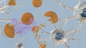
Cultivation of hMSCs on microcarriers
Previously, the authors demonstrated the feasibility of microcarrier-based cell expansion using a 3-L (Mobius 3 L, MilliporeSigma) single-use bioreactors to expand hMSCs that remained multipotent and continued to display both the genotypic and phenotypic characteristics of cells produced using traditional planar culture (2). A subsequent study described expansion of hMSCs in a 50-L (Mobius 50 L, MilliporeSigma) stirred tank bioreactor with refined control parameters and an improved media formulation after process development at 3-L scale (3).
The bioreactor cultures were monitored by cell counting, and also visualized at key time points by various staining methods for qualitative assessment of cell expansion and the availability of microcarrier surface area for growth. Good cell attachment to collagen-coated microcarriers was observed under constant agitation, pH, and dissolved oxygen control. For example, in the 50-L study there was an increase in the number of attached cells from 13% to 100% between 4 and 24 hours post-inoculation, with more microcarriers populated by one or more cells after 24 hours. Although bare microcarriers were observed in the culture at day three by staining of nuclei, fresh microcarriers were added at this time point in anticipation of increasing cell mass. By day 10 of the culture, almost all microcarriers were populated, showing that hMSCs can transfer to freshly-added microcarriers as their surface area requirement increases.
The migration of adherence-dependent cells from confluent to vacant microcarriers is critical to leverage a volume-up strategy for cell expansion in stirred-tank reactors. The 50-L bioreactor used in these studies has a 5:1 turndown ratio, meaning cultures may be inoculated at 10 L with volume-up feeds containing media and microcarriers added at key time points until the maximum operating volume of 50 L is reached. This approach allows for the addition of culture surface area and nutrients as cells expand without the necessity to detach cells and passage into new vessels, a distinct advantage over traditional planar culture formats.
Detachment from microcarriers
Cultivation of adherence-dependent cells such as Madin-Darby canine kidney epithelial cells (MDCK), Vero, or certain Chinese hamster ovary (CHO) cell lines on microcarriers has been used for vaccine and antibody production. Because the final therapeutic in many of these instances is secreted by cells into the culture medium, detachment and separation of cells from microcarriers are not required. In contrast, cells for use in regenerative medicine applications must be safely and effectively detached from microcarriers prior to further processing in a limited number of downstream steps. The culture surface properties of microcarriers can greatly impact the ability to achieve this detachment. An extracellular matrix (ECM) component coating or surface charge aids in attachment of the cells, which in turn deposit their own ECM as they expand on the culture surface. Growth factors commonly present in culture media have been shown to alter ECM deposition by hMSCs (4), and thereby may impact not only attachment efficiency, but also the cell-microcarrier detachment process. Downstream harvest conditions, therefore, should be considered while establishing upstream parameters, including media formulation and microcarrier selection.
Figure 1A: hMSCs were cultured on microcarriers in DMEM/10% FBS. Confluent microcarriers were incubated with the indicated concentrations of CellPrime rTrypsin for 15 min in order to determine the minimal effective concentration required for detachment of hMSCs. Fifteen percent of DMEM/FBS carry-over was maintained during phosphate buffer saline wash and detachment in order to model the bioreactor process.
The release of cells cultured on microcarriers can be accomplished by various techniques including trypsin proteolysis, cationic ion chelation, pH flux, application of hydrodynamic forces, or by some combination of these approaches (5). Detachment reagent concentration and detachment time must be carefully optimized to enable the maximum recovery of cells while preserving the quality of hMSCs. Results of a study using recombinant trypsin (rTrypsin) for detachment of hMSCs from microcarriers are shown in Figure 1. The concentration of rTrypsin on a per-million cells basis was titrated to determine the minimal amount of enzyme required to detach hMSCs from collagen-coated microcarriers, while maintaining high viability (Figure 1A). Incorporation of a trypsin-specific inhibitor into the process is particularly important for serum-free media processes as these formulations do not contain inherent inhibitors that are present in serum. High quality, plant-derived inhibitors are available that effectively quench enzymatic activity and can be used to stop the detachment step in a precise manner (Figure 1B).
Figure 1B: The effective concentrations of two animal origin-free trypsin inhibitors were determined by pre-incubating rTrypsin with the inhibitors at the indicated concentrations prior to detachment of hMSCs from culture flasks. hMSCs=human mesenchymal stem/stromal cells, FBS=fetal bovine serum, DMEM=Dulbecco’s Modified Eagle’s Medium.
Bioreactor systems with a raised harvest line facilitate performing the detachment process within the reactor tank that provides precise temperature and agitation control. At the end of culture, the impeller is stopped and microcarriers are allowed to settle to the bottom of the reactor tank. Spent media can then be drained via the side harvest line that resides higher than the bed volume of the settled microcarriers. The microcarriers may be washed with buffer prior to the addition of detachment enzyme. Alternatively, as in the case of serum-free media such as platelet lysate-supplemented formulations, concentrated enzyme may be added directly to the culture. Agitation is applied to expedite the detachment process and to leverage the bioreactor monitoring and control capabilities.
Separation of cells and microcarriers
Several technologies are available for the separation of cells from their expansion surface. Whereas enzymatic detachment alone is sufficient to separate cells from a planar culture system, cells grown in suspension require the development of methodologies to ensure efficient separation of cells from microcarriers. These culture beads are larger than hMSCs (generally approximately 150–200 µm compared with 15–20 µm, respectively) and can be separated from cells via size-exclusion filtration after the cells are detached. For this process, the authors created a normal flow filtration device that utilizes a woven mesh with a variety of sizes ranging from 40–100 µm to separate hMSCs from microcarriers. After the detachment of cells from microcarriers within the bioreactor, the microcarrier retention filter is welded onto the bottom harvest line in order to capture the full contents of the reactor. While cells pass through the filter, microcarriers are held back within the device. This approach has been successfully incorporated into both the 3-L and 50-L hMSC expansion processes (Figure 2) for the effective recovery of more than 500 million and 12 billion cells, respectively.
Figure 2: Process for harvest of hMSC expanded in stirred-tank bioreactors. Microcarrier-based culture in stirred tank bioreactors offers a high density alternative to planar culture in multilayer flasks. A closed system can be achieved by incorporation of single-use components including feed bags, sterile connectors, and the bioreactor itself. The harvest step, depicted here, can be accomplished by performing the cell-microcarrier separation step in the bioreactor, followed by retention of microcarriers using a normal flow filtration device, and collection of cells for further processing. Utilizing this approach in a 3-L bioreactor (Mobius 3 L by MilliporeSigma), hMSCs were effectively recovered and separated from microcarriers with >95% recovery when soy-derived trypsin inhibitors were used. The process has been applied successfully in 50-L hMSC expansion runs (3). hMSCs=human mesenchymal stem/stromal cells, PBS=phosphate buffer saline, FBS=fetal bovine serum.
Viability, cell-surface characteristics, and functional attributes are commonly assessed following the detachment and harvest steps to ensure quality has been maintained. A high recovery of hMSCs has been achieved using the microcarrier retention filter (e.g., 94% recovery of total cells with 99% cell viability at 3-L scale), with retention of cellular characteristics such as surface marker expression (CD105, CD90, CD73, and CD44, data not shown) comparable to the same hMSCs expanded in planar cultures. Because many clinical trials for hMSC therapies are investigating indications that involve immune system regulation, assays measuring hMSC inhibition of Tcell proliferation and the induction of indoleamine 2,3âdioxygenase were also used to evaluate cells harvested with the microcarrier retention filter after 50-L bioreactor expansion (3). No differences were detected between hMSCs cultured in planar culture and those from the bioreactor, highlighting the ability of the large-scale hMSC expansion and harvest process to yield highly functional hMSCs.
Conclusion
As cell-based therapeutics progress through clinical testing, there will be an increased need for scalable cell-manufacturing technologies that use non-animal-derived materials and are compatible with a limited number of downstream processing steps. Bioreactor-based manufacturing processes for hMSCs can drive higher cell yields per volume and unit operation, yet also introduce the requirement to separate cells from microcarriers. The incorporation of appropriately sized woven mesh filters or membrane filters into a normal flow filtration device is a simple and cost-effective single-use solution for the scales (3–200 L) required in regenerative medicine applications.
References
1. A. Schnitzler, Biochem. Bioeng. J. 108, 3–13 (2016).
2. D. Jing et al., BioPharm. Int. 26 (4), 28–38 (2013).
3. T. Lawson et al., Biochem. Bioeng. J. 120, 49-62 (Apr. 15, 2017).
4. S. Maxson et al., Stem Cells Transl. Med. 1 (2), 142-149 (February 2012).
5. A.W. Nienow et al., Biochem. Eng. J. 85, 79–88 (Apr. 15, 2014).
Newsletter
Stay at the forefront of biopharmaceutical innovation—subscribe to BioPharm International for expert insights on drug development, manufacturing, compliance, and more.
Advertisement
Related Content
Advertisement


Advertisement
Advertisement
Trending on BioPharm International
1
First-in-Human Study Validates Safety of Next-Generation mRNA–LNP Platform
2
FDA Clears PharmaResearch IND for Nano-Based Cancer Drug PRD-101
3
How CDMO Alliances Can Provide End-to-End Service that Reduces Drug Development Time and Costs
4
High-Dose Nusinersen Slows Neurodegeneration in SMA Patients, Study Shows
5