|Articles|April 1, 2013
- BioPharm International-04-01-2013
- Volume 26
- Issue 4
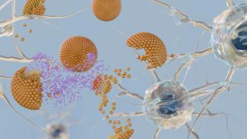
Growth Kinetics of Human Mesenchymal Stem Cells in a 3-L Single-Use, Stirred-Tank Bioreactor
The authors describe the growth characteristics of human mesenchymal stem cells cultured in a stirred-tank bioreactor.
Advertisement
Human mesenchymal stem cells (hMSCs), also known as marrow stromal cells, are a self-renewing population of adherent, multipotent progenitor cells with the capacity to differentiate into several cell lineages (1). In defined in vitro assays, hMSCs have been shown to readily differentiate into lineage-specific cells that form bone, cartilage, fat, tendon, and muscle tissues (1, 2). Mesenchymal stem cells also provide support and maintenance for the other major stem cell population in the bone marrow, the hematopoietic stem cells (2).
Human mesenchymal stem cells hold great promise as therapeutic agents because of their differentiation ability (thus their potential to replace damaged tissue) and for their immunomodulatory properties. A large number of clinical trials are underway that are using hMSCs in a variety of indications, including bone/cartilage disease, cancer, heart disease, gastrointestinal disease, diabetes, autoimmunity, and neurodegenerative diseases; hMSCs are also being used in drug discovery as a replacement for primary cells and animal models for initial toxicity and effector function screening of new compounds (3, 4). However, a key challenge remains for both drug discovery and clinical applications: obtaining a sufficient number of cells at reasonable cost (5).
The large-scale, industrialized production of hMSCs is necessary to advance these cells into clinical trials and to deliver the large quantities needed for drug discovery screening and lead optimization. Bridging the gap between basic research and large-scale manufacturing of stem cells for clinical trials requires the expansion of well-characterized cells produced under tightly controlled, consistent, reproducible culture conditions that adhere to cGMP standards. cGMP stem-cell culture systems require well-defined, optimized processes that support stem-cell expansion and differentiation to ensure consistent cell populations with uniform properties and predictable behaviors. Additionally, vessels used for expansion must allow rapid analysis of small volumes containing the actual cells to confirm that the expansion and harvest methods are yielding the expected cell populations. Because stem cells are the product, the sample size must be small enough to ensure that valuable product is not wasted.
Human mesenchymal stem cells have historically been isolated based on the ability of these cells to form adherent cell layers in culture and the concomitant lack of adherence of other cells in the bone marrow stroma, such as hematopoietic stem cells, adipocytes, and macrophages (1). However, the multilayer flatbed culture (2D) methods currently used for stem-cell expansion are cumbersome, time-consuming, do not allow for constant monitoring of cell characteristics throughout the expansion process, and they introduce a high degree of variability. These limitations make this method suboptimal for the manufacturing of clinical-grade hMSCs. Furthermore, the culture protocols for multilayer vessels require high labor cost, which in turn results in high cost of goods overall. Thus, the development of culture conditions that can be monitored and that produce high numbers of stem cells at low cost is warranted.
One possible solution for overcoming the limitations of 2D multilayer flatbed culture methods is the use of stirred tank bioreactors in which the stem cells are grown on a microcarrier scaffold for suspension (3D). In these 3D cultures, cell samples and medium can be analyzed throughout the expansion process and the growth process can be tightly controlled (e.g., oxygen, pH, glucose, glutamine, lactate, and ammonia). This article desribes the utility of EMD Millipore's 3-L single-use, bench-scale bioreactor (Mobius CellReady bioreactor, EMD Millipore, Billerica, Mass.) for the expansion of hMSCs.
MATERIALS AND METHODS
Isolation of hMSCs from unprocessed, fresh bone marrow
Unprocessed, fresh human bone marrows were purchased from Lonza (Walkersville, Md.). Upon receipt, unprocessed bone marrows were divided between 50-mL conical tubes with addition of an equal volume of phosphate-buffered saline (PBS, EMD Millipore). After spinning down at 1500 rpm for 5 min, supernatants were removed. Twenty-five mL of Red Blood Cell Lysing Buffer (Sigma-Aldrich, St. Louis, Mo.) was added and tubes were incubated on ice for 3 min. Cells plus lysis buffer were spun down at 1500 rpm for 5 min and supernatant was removed. After three cycles of lysis and spinning, cells were resuspended in 10% fetal bovine serum/Dulbecco's modified Eagle's medium (FBS/DMEM, Life Technology, Carlsbad, Calif.) and seeded at 10×106 cells/cm2 onto tissue-culture treated flasks. To calculate cumulative population doubling (cPD) by counting colonies, cells were placed at 1-, 10-, and 100×103 cells/cm2 in treated tissue-culture flasks.
Human mesenchymal stem-cell culture
In-house isolated hMSCs were cultured on 0.1% gelatin-coated (EMD Millipore) tissue-culture dishes in 5% CO2/95% air at 37 °C as described previously (1). The culture medium consisted of DMEM (Life Technologies) supplemented with 10% MSC-screened FBS (Life Technologies), 2 mM L-glutamine (EMD Millipore), 1× EmbryoMax Penicillin-Streptomycin Solution (EMD Millipore), and 8 ng/mL human recombinant basic fibroblast growth factor (bFGF, EMD Millipore). Cells were fed every other day by changing 100% of the medium. To protect hMSCs in bioreactors, medium was supplemented with 0.02% Pluronic-F68 surfactant (Sigma-Aldrich) and 37.5 ppm anti-foam C emulsion (Sigma-Aldrich).
For passaging, the cells were incubated with Trypsin-EDTA (EMD Millipore) in PBS and cell clumps were dissociated by gentle pipetting. The cell suspension was spun down at 200×g for 5 min, and after removing the supernatant, the cell pellet was resuspended in fresh medium and plated on tissue culture flasks at 3000 cells/cm2.
Preparation of microcarriers
Cytodex 1 and 3 (GE Healthcare, Piscataway, N.J.), collagen-coated and Hillix microcarriers (Solohill, Ann Arbor, Mich.), Cultispher G and S (Sigma-Aldrich) were prepared according to the manufacturer's instructions, including washing with PBS, sterilization by autoclave, and washing again with culture medium.
hMSC expansion on micro-carriers under static conditions
To evaluate the most suitable microcarrier for hMSCs culture in bioreactor, six different microcarriers from three vendors were tested. In particular, Cytodex 1 and 3 (GE Healthcare), collagen-coated and Hillix microcarriers (Solohill), Cultispher G and S (Sigma-Aldrich) were used for screening.
An equivalent total surface area of 250 cm2 and the same amount of hMSCs were used for each microcarrier in 10-cm low adherent petri dishes. Cell attachment efficiency was measured 24 hr after inoculation by calculating the ratio of the number of cells attached on microcarriers to the number of seeded cells. After 6 d of culture, cells were trypsinized and harvested from the beads. The cell release rate was calculated by the ratio of number of cells recovered from the beads to the total number of cells on the beads measured by NucleoCounter NC100 (New Brunswick Scientific, Enfield, Ct.).
Bioreactor inoculation, culture, and sampling
hMSCs just thawed or harvested from tissue-culture flasks were seeded directly into the Mobius CellReady 3-L Bioreactor (EMD Millipore) with microcarriers. To help cells attach to microcarriers, the agitation was set low, at around 25–35 rpm. The temperature of the bioreactor was set at 37 °C and supplemented with 5% CO2 from the bioreactor overlay.
Samples were withdrawn from bioreactors daily except on weekends. Cells and microcarriers were spun down at 200 rpm for 5 min and cell numbers were measured using a NucleoCounter NC100 (New Brunswick Scientific). Supernatants were collected and read by BioProfile FLEX (Nova Biomedical, Waltham, Mass.) for metabolic profile analysis.
hMSC differentiation to adipocytes and osteocytes
Mesenchymal stem cells cultured in bioreactors were trypsinized off the microcarriers and plated on 24-well tissue culture plates at seeding densities of 2.1×10⁴ cells/cm2 for inducing adipogenesis and at 4.2×10³ cells/cm2 for inducing osteogenesis. The induction of differentiation was carried out as per the instructions provided with the human mesenchymal stem cell functional identification kit (R&D Systems, Minneapolis, Minn.). After 21 d in culture, the cells were fixed and stained with Oil red O (EMD Millipore) to confirm the differentiation to adipocytes or with Alizarin red (EMD Millipore) to confirm the differentiation to osteocytes. Also, RNA was isolated from these differentiated cells to look at expression levels of differentiation markers using real-time polymerase chain reaction (RT-PCR).
RNA isolation, cDNA synthesis and RT–PCR
RNA was isolated from mesenchymal stem cells using RNeasy Mini Kit (Qiagen, Valencia, Calif.) as per the instructions in the protocol. 1µg total RNA was used for cDNA synthesis using qScript cDNA SuperMix (Quanta Biosciences, Gaithersburg, Md.). Real-time–polymerase chain reaction (RT–PCR) to quantify relative gene expression was carried out using custom PCR Array plates (SABiosciences, Valencia, Calif.) using the CFX96 Touch Real-time PCR system (Bio-Rad, Hercules, CA). PCR reaction was done with PerfeCTa SYBR Green SuperMix (Quanta Biosciences) as per the following cycling conditions: Initial denaturation and activation of AccuStart Taq DNA polymerase at 95°C for 10 min; PCR cycling for 40 cycles at 95 °C for 15 s and 60 °C for 1 min. The specificity of the amplification product was assessed by the melt curve method. The ΔΔCT method was used to quantify the relative gene expression by normalizing to the house-keeping gene.
Fluorescence-activated cell sorting
Cell pellets were resuspended in DMEM (Life Technologies) plus 2% BSA (Sigma-Aldrich) to bring the cell concentration to 5×10⁵ cells/mL. Cells were aliquoted into a round bottomed 96-well plate and antibody diluted in PBS (Cellgro, Manassas, Va.) was added. The plate was incubated at room temperature protected from light for 20 min. PBS was added after incubation and the plate was centrifuged to pellet the cells. The supernatant was discarded and cells resuspended in PBS. The plate was read on a guava easyCyte 8HT Flow Cytometry System (EMD Millipore). Raw data were imported into FlowJo (Treestar, Ashland, Ore.) for analysis.
The antibody and isotype control were listed as following: CD 105-FITC, CD 73-PE (AbCam, Cambridge, Mass.), CD 90-FITC, CD 11b-PE, CD 79α-PE, CD 14-FITC, CD45-FITC, CD 34-FITC, CD 19-FITC, HLA-DR-FITC (EMD Millipore), CD 146-FITC, CD 44-FITC, CD 45-FITC, CD 106-FITC, CD 274-FITC (AbD Serotec, Raleigh, N.C.), IgG1k FITC, IgG2ak FITC (BD Bioscience, Sparks, Md.), IgG1 PE, IgG2a PE (R & D Systems,), and IgG2b FITC (AbD Serotec).
Karyotyping and StemArray assays
hMSCs harvested from bioreactors or CellStack were harvested and seeded at 3000 cells/cm2 in T25 flasks. Flasks were filled with fresh medium and shipped to Cell Line Genetics Inc. for characterization. Karyotyping, FISH, and StemArray assays were done according to their protocols.
Statistical analysis
Data are expressed as mean± standard deviation unless stated otherwise. Statistical analysis including ANOVA was performed using Minitab (Minitab Inc, State College, PA). P values less than 0.05 were considered as significant.
RESULTS AND DISCUSSION
The in-house derived hMSCs used throughout this study were first characterized through analysis of their cell surface antigen profiles and their differentiation ability toward adipogenic and osteogenic lineages (see Figure 1). These cells demonstrated the appropriate CD90+ , CD105+ , CD73+ , CD34- , CD45- , CD11b- cell surface antigen profile and the ability to differentiate toward all three lineages.
Figure 1: (a) In-house derived human mesenchymal stem cells (hMSCs) express CD90+, CD105+, CD73+, CD34-, CD45-, CD11b- cell surface antigen profiles; (b) In-house derived hMSCs retain differentiation ability toward adipogenic, osteogenic and chondrogenic lineages.
For 3D hMSC expansion, cells can either be expanded first on T-flasks, or thawed and directly seeded in the Mobius CellReady 3-L Bioreactor on microcarriers (see Figure 2). To determine the best parameters for 3D expansion, various cell and microcarrier seeding densities were investigated (see Figure 3). Optimal cell seeding density was demonstrated at 5 million cells/L and optimal microcarrier seeding density was demonstrated at a concentration of 15 g/L.
Figure 2: (a) Human mesenchymal stem cells (hMSCs) can be either expanded first on T-flasks or thawed and directly seeded into a Mobius CellReady 3-L bioreactor with microcarriers; (b) Morphology of hMSCs on microcarriers after expansion in the bioreactor (Upper: CellTracker Green; Lower: bright field).
To study the effects of aeration on the growth of hMSCs in the bioreactor, 5 million cells/L were seeded per bioreactor, allowed to grow for 6 d, and then various sparge strategies were executed, including aeration through minisparge and open-tube at both high and low air-flow rates (see Figure 4). By day 12, cell growth reached a maximum in the control bioreactor, which could either be due to shear force by air bubbles or a preferred lower oxygen culture.
Figure 3: (a) Human mesenchymal stem cell (hMSC) attachment affinity 24 h; (b) Microcarrier concentration of 15 g/L demonstrates maximum hMSC expansion after 11 d.
The growth rate of hMSCs was then compared in 2D and 3D cultures (see Figure 5). hMSCs not only proliferated on microcarriers in the bioreactor, but also colonized empty microcarriers. After a one to three day lag phase, hMSCs expanded quickly in the bioreactor and reached maximum cell number (approximately 600 million cells) in 12 d, which is roughly 50% greater than expected from growth in 2D culture after 14 d. Flow cytometry data illustrated no difference in cell surface antigen expression between hMSCs expanded via 2D or 3D culture.
Figure 4: (a) Sparge strategies; (b) Cell number reached a maximum in the control bioreactor; (C) dO2 level remained fairly constant except in the control bioreactor.
hMSCs were characterized following expansion for two weeks in 2D and 3D cultures (see Figure 6). Expanded cells were exposed to differentiation media toward adipogenic and osteogenic lineages, cytogenetic analysis, and cell functional analysis. Regardless of method of expansion, expanded hMSCs demonstrated multipotent differentiation abilities, normal male karyotype, and the ability to secrete important cytokines, including interferon-gamma, interleukin-6 and interleukin-8.
Figure 5: (a) Human mesenchymal stem cells (hMSCs) not only proliferated on initial seeded microcarriers but also colonized empty microcarriers; (b) hMSCs expanded quickly in the bioreactor and reached maximum cell number (~600 million cells) in 12 d. The grey bar represents the expected cell number from a 10-Stack CellSTACK in 14 d; (C) No difference in cell surface antigen expression between hMSCs expanded in CellSTACK and a bioreactor with microcarriers (dotted line: isotype control; dark blue line: bioreactor; light blue line: CellSTACK).
Spent media (glutamine, glucose, lactic acid and NH4+) from 2D and 3D culture were analyzed and compared (see Figure 7). Additionally, multiple parameters that potentially affect hMSC expansion in 3D cultures were examined, including the concentration of lactic acid in the media and feeding strategies (see Figure 7). The capacity to monitor and control the metabolism of cells in a bioreactor facilitates a feeding strategy to maximize cell number.
Figure 6: (a) After expansion for two weeks in CellSTACK and Mobius CellReady 3-L bioreactor, human mesenchymal stem cells (hMSCs) demonstrate multipotent differentiation abilities; (b) Expanded hMSCs retain normal male karyotype; (C) Induced expanded hMSCs secrete important cytokines, including IFN-γ, IL-6, and IL-8.
The effect of lactate and glucose concentration and pH on the expansion of hMSC on microcarriers in suspension culture was tested (data not shown). hMSCs were able to grow under conditions of no glucose by consuming other nutrients in the medium; however, the growth rate was reduced. hMSCs were able to expand under conditions of high glucose concentration (4.5 g/L) without significantly reducing growth rate. High concentrations of lactate (> 1/6 g/L) inhibit hMSC growth on microcarriers, which is consistent with 2D culture results. Growth was significantly reduced at low pH (< 6.8).
Figure 7: (a) Spent media (glutamine, glucose, lactic acid and NH4+) from bioreactor and CellSTACK were analyzed; (b) Parameters (e.g., concentration of lactic acid in the media, dO2, pH and feeding strategies) that affect hMSCs expansion in bioreactor were studied.
CONCLUSION
Taken together, these data demonstrate that expansion of hMSCs in 3D culture demonstrate similar flow cytometry profiles, differentiation ability, and functional status as hMSCs expanded in 2D culture. After two weeks, cells reach densities greater than 2×10⁵ cells/mL while maintaining their identity as shown by the surface expression of CD105, CD90, and CD73 and the absence of CD14, CD34, and CD45.
The production of well-characterized hMSCs in stirred-tank bioreactors using microcarriers is the first step towards establishing a scalable, single-use production process for the large-scale industrialization of stem cells. The Mobius CellReady 3-L bioreactor presents several advantages compared with multilayer flatbed cultures, including lower overall cost, ease of use, and the ability to closely monitor process parameters through-out expansion and make adjustments while cells are still viable, if needed.
Donghui Jing, PhD*, is a research scientist III, Stem Cell Initiative at EMD Millipore; Neethu Sunil is a research scientist at Experis, Southborough, Mass. Sandhya Punreddy is a research scientist III; Manjula Aysola, PhD, is a research scientist III; Daniel Kehoe, PhD, is a research scientist III; Julie Murrel, PhD, is a senior scientist; Martha Rook, PhD, is head of upstream and sensor technologies; and Knut Niss, PhD, is R&D manager; all at EMD Millipore, Bedford, Mass.
*To whom correspondence should be addressed,
REFERENCES
1. D.J. Prockop, Science 276, 71-74 (1997).
2. M.F. Pittenger and D.R. Marshak, in Stem Cell Biology, D. R. Marshak, R. L. Gardner, and D. Gottlieb, Eds., (Cold Spring Harbor Laboratory Press, Cold Spring Harbor, New York, 2001).
3. D. Jing et al., Tissue Eng Part B Rev. 14 (4): 393-406 (2008).
4. S.S. Kitambi and Chandrasekar, Stem Cells and Cloning: Advances and Applications 4, 51-59 (2011).
5. J. Rowley et al., BioProcess Intl. 2012 Mar; 10 (S3), 16-22 (2012).
Articles in this issue
almost 13 years ago
Milestones and Moderate Progress in 2012 Drug Approvalsalmost 13 years ago
Custom-Cell Products: Diversified In Vitro Modelsalmost 13 years ago
Benchmarking: Don't Just Run the Numbers, Understand the Processalmost 13 years ago
Strategic Partnering for Manufacturingalmost 13 years ago
State of Quality and Compliance in the Biopharmaceutical Industryalmost 13 years ago
Navigating Emerging Markets — Latin Americaalmost 13 years ago
Analytical Method Lifecycle: A Roadmap for Biopharmaceutical Developmentalmost 13 years ago
Manufacturers Under Pressure to Manage Painkillersalmost 13 years ago
Prescribing Caution for Biosimilarsalmost 13 years ago
Financing InnovationNewsletter
Stay at the forefront of biopharmaceutical innovation—subscribe to BioPharm International for expert insights on drug development, manufacturing, compliance, and more.
Advertisement
Related Content
Advertisement


Advertisement
Advertisement
Trending on BioPharm International
1
First-in-Human Study Validates Safety of Next-Generation mRNA–LNP Platform
2
FDA Clears PharmaResearch IND for Nano-Based Cancer Drug PRD-101
3
How CDMO Alliances Can Provide End-to-End Service that Reduces Drug Development Time and Costs
4
High-Dose Nusinersen Slows Neurodegeneration in SMA Patients, Study Shows
5