- BioPharm International-03-01-2015
- Volume 28
- Issue 3
Detecting Protein Aggregates and Evaluating their Immunogenicity
The thorough analysis of a therapeutic protein product’s propensity to aggregate may be a necessary step in the prevention of a cell-mediated immune response.
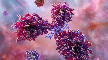
Aggregation in biopharmaceuticals remains a major concern and threatens the stability of a product. Protein aggregation can occur during all stages of the lifetime of a protein therapeutic, including expression, refolding, purification, sterilization, shipping, storage, and delivery processes (1). The mechanism of protein aggregation is not well understood, “but most proteins will aggregate when they are in an unfolded or partially folded state,” says Angelica Olcott, product manager at ProteinSimple. Aggregation typically occurs because of nucleation, surface-induced factors, as a result of conformationally or chemically altered monomers, or association of native monomers (2). When a protein has both positive and negative charges on its surface, protein
protein electrostatic interactions are highly probable, and these can lead to aggregation (2). The very nature of aggregation is challenging, says St John Skilton, senior global market manager of biologics at SCIEX, a life-science analytical technologies company. The fact that aggregation is “dynamic, that it is a mixture in solution, and that species do not attach to each other permanently, but through bonds that can be broken by ionic processes, means that we need to rely on both separation and detection,” which makes it especially difficult.
Aggregation risk factors
Certain manufacturing stages influence the risk of chemical degradation, which increases the risk of physical degradation and the formation of aggregates. “These stages include formulation composition, presence of microbial or vial contaminants during cell culture, and storage,” notes Olcott. Work by Hasija et al. (2) on forced degradation studies in vaccines have helped evaluate the propensity of a product to aggregate at various points in the manufacturing process.
Higher concentrations of a protein formulation can increase the probability of aggregation, as well as changes in solution pH, temperature, buffer choice, agitation, and storage conditions. “Although components of the formulation are designed to preserve protein conformation, sometimes these excipients (e.g., human serum albumin, polysorbate) can interact with the protein and destabilize it,” notes Olcott. “The storage container environment plays a role too; studies in prefilled syringes show that silicone oil, leachables from the rubber stopper, and glass delamination can induce aggregates.” Denaturation and degradation of therapeutic proteins during storage, then, must be carefully considered, and testing for extractables and leachables should be performed under all conditions. “Because it is largely impossible to predict aggregation, it’s important to monitor it consistently during the specific events which are known to cause stress on the formulation,” comments Olcott.
While many analysis instruments can measure particle size distribution, to identify the nature of particulates in protein therapeutics, information about particle shape and morphology is necessary, says Olcott. This information will help investigators distinguish between extrinsic particules (foreign matter), inherent particles (protein aggregates), and intrinsic particles (such as silicone droplets), according to Lew Brown, technical director at Fluid Imaging Technologies. New technologies for protein characterization tell us more about particle classification, as opposed to simply providing particle counts. Regulatory agencies are increasingly asking drug manufacturers to characterize particulate matter in biologics.
Unfortunately, there is little general agreement about how to handle the problem of aggregation, according to many experts. Brown notes there does not seem to be a consensus yet in whether the presence of submicron (<1μm) aggregates can even be used as a predictor of whether subvisible (2–100 μm) aggregates will form. Different techniques and particle-detection methods for measuring aggregation at various sizes produce different results that may or may not correlate with one another. “All of this is caused by the difficulties in accurately measuring protein aggregation in repeatable experiments,” Brown comments.
Particle-detection methods
There are various detection technologies that should be used to address protein aggregation needs at various phases of the biopharmaceutical development process; however, no single analytical method alone can provide an accurate representation of protein masses. Different methods apply to different classes of aggregates, says Daniel Some, PhD, director of marketing and principal scientist at Wyatt Technology Corporation, and these classes include reversible vs. irreversible aggregates and small (or soluble) vs. large (or insoluble) aggregates.
To measure across size range, Olcott insists one needs a variety of methods. “Protein aggregates in the micron range can be quantified by several analytical techniques, including light obscuration (LO), dynamic imaging particle analysis (DIPA) techniques such as micro-flow imaging (MFI), and Coulter counter (CC). Each of these methods measures different characteristics, but all three will report particle size and count. MFI differs from LO and CC by also providing particle shape and image intensity, for particles from 1–300 microns,” she adds.
LO alone is not sufficient to detect aggregates, as it is not sensitive enough to always capture translucent particles. Aggregation can cause protein particles to become transparent, and relying solely on LO can lead to undercounting, notes Olcott. Additionally, as Brown points out, LO “can only measure particle size via an indirect measurement of volume converted to size based on assumption of spherical shape,” so imaging systems must also be used to capture amorphous particles.
Proteins can be distinguished from other particles through tools such as MFI, which shows that silicone oil microdroplets appear very different from protein aggregates in formulation, says Olcott. With MFI, researchers can now determine how particle groups vary in their samples, and with “new high-throughput screening software, users can instantly compare results across many samples,” notes Olcott.
Another way to measure the presence of aggregation is mass spectrometry. “Under the right conditions, mass spectrometry is particularly useful for structural characterization of intact protein aggregates when quarternary protein structures can be maintained,” Skilton notes. When performing an analysis by mass spectrometry, SCIEX can use electrospray, allowing a plume of ions to enter the mass spectrometer. This technique helps link liquid chromatography or capillary electrophoresis electrospray (CESI) easily and allows orthogonal techniques to work together for better information content. With peptide mapping, investigators can choose from “a panoply” of enzymes to perform digestions at different locations and review the information in software programs to determine aspects of higher-order structure. “Linking capillary electrophoresis with mass spectometry brings additional power to peptide mapping by improving ionization efficiencies,” Skilton says. This information can help investigators locate expected disulfide bonds, which maintain the higher-order structure of a protein, he adds.
Let there be light
For submicron aggregates, investigators routinely use size-exclusion chromatography (SEC), which Olcott says separates particles according to hydrodynamic size. Users can add a light-scattering detector in line after SEC for irreversible aggregates, notes Some, and batch (unfractionated) light scattering methods can be applied to reversible aggregates. Analytical ultracentrifugation (AUC) and asymmetrical-flow field-flow fractionation (AF4) are orthogonal methods to SEC, and are thought to provide a thorough picture of aggregate quantitation (3).
Some notes that small, irreversible aggregates in the range of oligomers are best quantified by multi-angle static light scattering (MALS) coupled with SEC (SEC–MALS). “This method provides absolute determination of molar mass, independently of SEC column calibration, so we can be certain of the identification of eluting species.” Larger, irreversible aggregates of “up to hundreds of nm in radius” are best characterized by field-flow fractionation multi-light scattering (FFF–MALS), Some comments. “FFF separates macromolecules and nanoparticles with no stationary phase, and the addition of MALS detection provides for determination of size and molar mass.”
“Batch dynamic light scattering (DLS) is a useful tool to characterize aggregates when the dilution or shear encountered in SEC or FFF causes the aggregates to disassociate or when aggregation is to be measured under many conditions and/or temperatures,” says Some. Though DLS does not provide the high resolution of SEC–MALS or FFF–MALS, notes Some, it can be automated via a high-throughput plate reader. To characterize reversible, soluble aggregates that occur at high-protein concentrations, composition-gradient light scattering (CG–MALS) is best applied.
Because older technologies such as LO and CC cannot classify submicron-sized particles, this can limit their ability to screen for potential immunogenic risk, says Olcott. Nanoparticle tracking analysis and resonant mass measurement are new technologies that facilitate better particle classification for this purpose, says Brown, and aggregates are best measured through DIPA systems. These systems image the particles in a moving fluid stream and are able to “capture statistically significant amounts of data in a short time frame.”
Ripple writes that while submicron particle detection would benefit from improvements in quantitation, subvisible particle discovery requires better system standards for comparisons across different technologies (4). There is currently no statutory requirement for the presence of protein aggregates in the subvisible range, so the ultimate goal is “protein stability with the lowest possible level of aggregates,” asserts Olcott.
When other methods are not feasible or when direct assessment of the aggregation in a sampled vial is required, AUC is frequently used, says Randall Lockner, strategic marketing manager for centrifugation at Beckman Coulter Life Sciences. According to Lockner, with AUC, the amount of method development needed is minimal, as biopharmaceuticals can often be analyzed in the formulation buffer as it exists in the vial. Because AUC can provide information about a sample, such as energetics, stoichiometry, conformation, concentration, shape, and solvation state, Lockner considers AUC “an indispensible orthogonal method in biopharma as a primary, first-principles technique used to validate other analytical techniques.”
Different instruments will provide a scientist with different results, however, because they rely on different measurement principles, and they can often produce conflicting results. “Because of this variability in measurements using different techniques, it is usually necessary to use multiple, orthogonal techniques,” says Brown. According to Brown, there are no standard reference materials with which to compare proteinaceous materials, and as a result, no verifiable techniques with which to quantify aggregates. “It seems that the majority of fluctuation on measurements is introduced by not having a very consistent, highly detailed [standard of procedures] for making measurements.” The consensus among the experts is that further analytical headway is needed to better classify and characterize the diversity of particles that investigators encounter. “Bearing this challenge in mind, NIST [The National Institute of Standards] recently completed a round-robin study of particle analysis technologies at 23 different pharma and academic sites with an investigational protein-like standard called ETFE [ethylene tetrafluoroethylene]” (5), says Olcott.
There are still some major shortcomings in available particle detection technology, however. Having a way to cross-compare results obtained from a variety of different technologies would be helpful, notes Olcott. There are still only a few technologies that span submicron to micron, and even those that are available are usually limited to particle size and can only handle small samples. “Being able to measure particle mass accurately and directly across the entire particle size range would be very beneficial,” she adds.
Immunogenicity and efficacy
High levels of aggregates can reduce therapeutic efficacy of a biologic, or impair product quality or stability. This could increase the risk of an immunogenic response, and according to Olcott, filtering the therapeutic product does not eliminate the risk of aggregates completely. “The presence of protein aggregates reduces the quantity of the active therapeutic protein available,” she says, and “in some rare instances, the protein aggregate can actually be super-potent compared with the stable biologic.” FDA, Guidance for Industry, Immunogenicity Assessment for Therapeutic Protein Products (6) notes that unwanted immune responses can also occur “by cross-reacting to an endogenous counterpart, leading to the loss of its physiological function.” The guidance document points to an example with pure red cell aplasia, where the induction of anti-erythropoietin antibodies neutralized the native protein. Minor changes in formulation, therefore, can lead to very serious adverse events, and conditions that could lead to an increased tendency for aggregation should be carefully monitored.
Specifically, FDA outlines in the aforementioned guidance that protein aggregates elicit an immune response in humans through the following mechanisms: by causing B-cell activation as a result of cross-linked B-cell receptors; by promoting antigen uptake, processing, and presentation; and by “triggering immunostimulatory danger signals.” These actions have the ability to recruit T cells and may play a role in neutralizing product efficacy (6).
Olcott points out that the potential causes of immunogenic response are difficult to measure and are dependent on the “cellular and humoral immunity in the intended patient population.” While assays for anti-drug antibodies and product-specific antibodies can be used to define immunogenic risk, and can be used throughout the specific dosing cycle, they can usually only tell investigators about patient-specific factors, such as allergy status and HLA (human leukocyte antigen) haplotype. “Unfortunately, animal models cannot be used to predict the human response to a therapeutic protein product,” adds Olcott. “However, they can show the consequences of an immune response, if using a species-specific version of a therapeutic protein.” FDA encourages manufacturers to explicate the underlying mechanism(s) of action for immunotherapies to mitigate the risk of clinical consequences (6).
Certain chemical modifications to a protein product-such as oxidation, deamidation, aldehyde modification, and deamination-can cause aggregates to form once a product is inside the body and could promote an immune response, even if these alterations do not cause product aggregation during manufacture or storage. Olcott notes that the effect of processes such as oxidation could make certain types of protein aggregates more immunogenic than others, and many particles could be a heterogenous mixture of protein aggregates with other contaminants, such as silicone oil. FDA notes that changes to protein chemistry that may otherwise be well controlled in other environments “may occur in vivo in the context of the relatively high pH of the in-vivo environment or in inflammatory environments and cause loss of activity as well as elicitation of immune responses.” Thus, FDA suggests therapeutic protein products be evaluated in the “in-vivo milieu in which they function” whenever possible to “facilitate product engineering to enhance the stability of the product under such stress conditions” (6).
To further reduce the risk of aggregates, FDA also recommends selection of an appropriate cell substrate, a facility that employs GMPs, a robust purification protocol, and formulation/container closure systems that do not preclude aggregation.
Size doesn’t matter
Protein aggregates of any size in therapeutic products can create an immunogenic response, a fact that studies have confirmed for half a century, notes Olcott. “No clear dose-response relationship exists between the quantity of aggregates and the resulting immunogenic response,” she says. In addition, there is no guarantee that if larger aggregates are filtered, the chance of an immune response decreases, although FDA claims there is some evidence that higher-molecular-weight aggregates can elicit a more powerful response (6). “So far, there does not seem to be any conclusive data directly relating submicron aggregates to immunogenicity,” Brown asserts. Steps can be taken to reduce aggregation regardless of aggregate size, such as modifying proteins through glycosylation or PEGylation, but product efficacy or dosage could change as a result. “Given the lack of clarity on human immunogenicity, recommendations should remain product-specific,” concludes Olcott. While there is still some disagreement about the significance of aggregates and their potential immunogenicity, as the demand for high-concentration protein formulations increases, says Lockner, “understanding aggregation in product formulations can be a key competitive advantage.”
References
1. E.Y. Chi,
2. M. Hasija, L. Li, and N. Rahman et al., Vaccine: Development and Therapy 3 1133 (2013).
3. R. Manning et al., “Review of Orthogonal Methods to SEC for Quantitation and Characterization of Protein Aggregates,” BioPharm International 27 (12) 2014.
4. D.C. Ripple and M.N. Dimitrova, J. Pharm. Sci. online, DOI: 10.1002/jps.23242, 2012.
5. D.C. Ripple, C.B. Montgomery, and Z. Hu, J. Pharm. Sci. 104 (2) 666-677 (November 2014).
6. FDA, Guidance for Industry: Immunogenicity Assessment for Therapeutic Protein Products (Rockville, MD, August 2014).
Article DetailsBioPharm International
Vol. 28, No. 3
Pages: 22-26
Citation: When referring to this article, please cite it as R. Hernandez, “Detecting Protein Aggregates and Evaluating their Immunogenicity,” BioPharm International 28 (3) 2015.
Articles in this issue
almost 11 years ago
USP Publishes Monoclonal Antibody Guidelinesalmost 11 years ago
Maintaining the Stability of Biologicsalmost 11 years ago
Springing Forwardalmost 11 years ago
Reinventing the Cold Chain in a High-Stakes Marketalmost 11 years ago
A Robust CAPA System for a Global Supply Chainalmost 11 years ago
CMOs Plan for Capacity Expansionsalmost 11 years ago
Vaccine Development and Production Challenges Manufacturersalmost 11 years ago
Re-use of Protein A Resin: Fouling and Economicsalmost 11 years ago
SEC in the Modern Downstream Purification Processalmost 11 years ago
Polyethylene Biocontainer Increases CustomizabilityNewsletter
Stay at the forefront of biopharmaceutical innovation—subscribe to BioPharm International for expert insights on drug development, manufacturing, compliance, and more.


